Systemic lupus erythematosus (SLE) is a heterogeneous, autoimmune, inflammatory, connective tissue disease affecting multiple organs. Neither central nervous system nor peripheral nervous systems are spared. The neurologic system is involved in a wide range of 10–80% of patients with SLE.
Peripheral neuropathy, such as Guillain-Barré syndrome (GBS) and its variants, can occur in neurologic complications of SLE. GBS variants include: 1) acute inflammatory demyelinating polyneuropathy (AIDP); 2) acute motor-sensory axonal neuropathy (AMSAN); 3) Miller Fisher syndrome (MFS), a rare variant with ocular involvement; and 4) acute motor axonal neuropathy (AMAN). Other rare variants can also occur.
Below, we present a case of an African-American woman with SLE presenting with the MFS variant of GBS. She completely recovered from MFS, which involved ophthalmoplegia, hyporeflexia and ataxia, with plasma exchanges and high-dose steroids. We also review the pathophysiology, treatment modalities and prognosis of this condition.
The Case

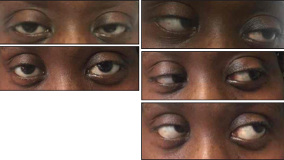
Top left: Ptosis of the left eye at presentation. Bottom left: Resolution of the ptosis after treatment. Top right: Left lateral rectus palsy at presentation. Middle right: Resolution of the left lateral rectus palsy, lateral gaze. Bottom right: Outward and upward gaze.
In early 2017, our rheumatology service was consulted for a 40-year-old African-American woman with complaints of double vision, bilateral lower-extremity numbness and ascending weakness with worsening over a two-month period. She was unable to walk without assistance on admission. A review of systems was positive for numbness in her lower legs ascending to her mid-chest, double vision, drooping of her left eyelid, one episode of diarrhea and difficulty walking.
Her medical history was also significant for hypertension. There was no history of intravenous drug use, tobacco use, HIV, hepatitis, clotting disorders or miscarriages.
She had a history of SLE with its onset in 2014, when it manifested with alopecia, mucosal ulcers, arthritis, elevated anti-nuclear antibodies (ANA), anti-double stranded DNA (anti-dsDNA), hypocomplementemia and anti-Smith (Sm) antibodies. She was placed on hydroxychloroquine 200 mg twice daily at that time.
The physical examination revealed a wide-based ataxic gait, a convergent gaze, left ptosis, left lateral rectus palsy (see Figure 1), horizontal diplopia, hyperpigmented lesions involving the antihielix/acoustic meatus, alopecia, hyporeflexia 0/5 in the ankles and knees bilaterally, 3/5 motor function for the left lower extremity and 4/5 for the right lower extremity. Her lower extremities had normal vibratory sensation below the level of the ankles. There was no clinical evidence of synovitis.
Miller Fisher syndrome (with its triad of ophthalmoplegia, ataxia & areflexia) is a rare variant of Guillain-Barré syndrome.
Laboratory analysis revealed an elevated erythrocyte sedimentation rate (ESR) of 127 mm/h, a C-reactive protein (CRP) level of 0.1 mg/L, hemoglobin of 10.9 mg/dL, hematocrit at 32.1%, hypocomplementemia (i.e., C3 at 44 and C4 at 9), ANA at 1:2560, anti-SSA antibody at 215, dsDNA at 1:640 and anti-Sm antibodies at 400. Creatinine phosphokinase (CPK), hepatitis panel, thyroid function tests, B12 and methylmalonic acid (MMA) were normal. Cerebrospinal fluid (CSF) protein was elevated at 72 mg/dL, with a normal white blood cell count of 2 mm3. There were no oligoclonal bands. The angiotensin converting enzyme (ACE), neuromyelitis optica (NMO) immunoglobulin G (IgG) antibody, paraneoplastic panel, lyme cerebrospinal fluid (CSF) and ganglioside GQ1b antibodies were negative.