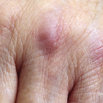
Several studies have looked at the most cost-effective and high-yield ways to screen for malignancy via imaging in patients with scleroderma or dermatomyositis. In a retrospective analysis of 400 patients with dermatomyositis, Leatham et al. identified 48 patients with a total of 53 dermatomyositis-associated cancers.5 Twenty-seven of these patients had an undiagnosed malignancy at the time of dermatomyositis diagnosis, and the majority (59%) of these cancers were asymptomatic and detected by computed tomography (CT). These data indicate that blind CT of the chest, abdomen and pelvis may be a high-yield approach to screening for malignancy in dermatomyositis.
In another study of 55 patients with newly diagnosed myositis, whole-body fluorodeoxyglucose-positron emission tomography/CT (FDG-PET/CT) was compared with the results of conventional cancer screening, which included thoracoabdominal CT, mammography, gynecologic examination, ultrasonography and tumor marker analysis. In this study, the positive and negative predictive values for diagnosing occult malignancy were 85.7% and 93.8%, respectively, for FDG-PET/CT and 77.8% and 95.7%, respectively for conventional cancer screening. From this, the authors concluded the performance of a single FDG-PET/CT for diagnosing occult malignancy in patients with myositis was comparable to that of broad conventional screening that involves many more tests.6
In Sum
In concluding her lecture, Dr. Shah urged the audience to think carefully about which patients with rheumatologic diseases are at increased risk of cancer and to use all tools at the clinician’s disposal, including personal and family history, physical exam, autoantibody profiles and clinical disease features to risk-stratify patients for cancer screening.
There may not be a one-size-fits-all model for such screening in rheumatic disease patients, but that is exactly why a thoughtful rheumatologist can be most helpful in guiding patients on this difficult subject.
Jason Liebowitz, MD, completed his fellowship in rheumatology at Johns Hopkins University, Baltimore, where he also earned his medical degree. He is currently in practice with Skylands Medical Group, N.J.
References
- Joseph CG, Darrah E, Shah AA, et al. Association of the autoimmune disease scleroderma with an immunologic response to cancer. Science. 2014 Jan 10;343(6167):152–157.
- Igusa T, Hummers LK, Visvanathan K, et al. Autoantibodies and scleroderma phenotype define subgroups at high-risk and low-risk for cancer. Ann Rheum Dis. 2018 Aug;77(8):1179–1186.
- Fiorentino DF, Chung LS, Christopher-Stine L, et al. Most patients with cancer-associated dermatomyositis have antibodies to nuclear matrix protein NXP-2 or transcription intermediary factor 1γ. Arthritis Rheum. 2013 Nov;65(11):2954–2962.
- Bernet LL, Lewis MA, Rieger KE, et al. Ovoid palatal patch in dermatomyositis: A novel finding associated with anti-TIF1γ (p155) antibodies. JAMA Dermatol. 2016 Sep1;152(9):1049–1051.
- Leatham H, Schadt C, Chisolm S, et al. Evidence supports blind screening for internal malignancy in dermatomyositis: Data from 2 large U.S. dermatology cohorts. Medicine (Baltimore). 2018 Jan;97(2):e9639.
- Selva-O’Callaghan A, Grau JM, Gámez-Cenzano C, et al. Conventional cancer screening versus PET/CT in dermatomyositis/polymyositis. Am J Med. 2010 Jun;123(6):558–562.


