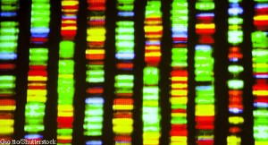
 Through gene analysis, researchers have found different types of interferons in systemic lupus erythematosus (SLE) tissues and cells, such as skin and synovium. The analysis, which probed 2,000 gene expression datasets from SLE patients, specifically investigated modules of genes derived from the downstream interferon gene signature. It found enriched downstream interferon signatures that were predominately from IFNB1. These interferon signatures were higher when compared with the expression of downstream interferon signatures in kidneys with lupus nephritis, according to the study, published April 23 in Nature Communications Biology.1
SLE is associated with the excessive production of interferons and autoantibodies against nucleic acids. Previous research has found the activation of the type I interferon system in SLE affects the innate and adaptive immune systems.2
This new investigation was led by Michelle D. Catalina, PhD, senior scientist of systems immunology, and colleagues at AMPEL BioSolutions LLC, Charlottesville, Va. The researchers used the specific and unique gene signatures of multiple interferons to better understand the roles of various interferons in lupus pathogenesis, as well as to gain insight into the appropriate use of interferon signature(s) as a possible biomarker.
These results add to previous findings that the interferon gene signature is readily detectable in patients with inactive disease and does not change synchronously with disease activity. Dr. Catalina says, “The interferon gene signature has been known to be associated with SLE for more than 15 years. There are, however, numerous interferon species, and it has remained unclear which of the various interferons is contributing to lupus pathogenesis.”3,4
Understanding the specific interferons involved in lupus and the roles of each in disease manifestation may provide essential information to safely and effectively target these molecules for treatment, she acknowledges. Example: It’s known interferons cannot only contribute to disease pathogenesis, but also are important in host defense against a variety of viral pathogens.
Type 1 & 2 Interferons
For this investigation, interferon gene signature types were identified in SLE blood samples, using gene set variation analysis with IFNA2, IFNB1, IFW1, IFNG, TNF, IL12 and interferon core signature genes. The goal was to determine the relative enrichment of these signatures in the whole blood or peripheral blood mononuclear cells of SLE patients and healthy controls.
Heatmap visualization of gene set variation analysis enrichment scores showed patients with highly enriched signatures for IFNA2, IFNB1, IFNW1, IFNG and the interferon core. Most SLE patients were separated from controls by these scores. In most SLE patients, the gene set variation analysis enrichment scores were the strongest for the type I interferons compared with IFNG, TNF or IL12. However, some SLE patients had no type 1 or type 2 interferon gene signatures, but did possess a TNF or IL-12 signature.
“This work demonstrated, unexpectedly, the predominance in both SLE blood and tissues of an [interferon] beta signature, whereas most previous efforts have assumed this signature was induced by [interferon] alpha,” Dr. Catalina says. “Also, our work clearly demonstrated, for the first time in adult subjects with SLE, the lack of a relationship between the presence of an interferon signature and disease activity. This [finding] is perhaps an explanation for the failure of anifrolumab to meet its primary endpoint—a change in disease activity.”
In further describing the study’s findings, Dr. Catalina says the data clearly show SLE patients with very low disease activity can have a robust interferon signature. The data also show interferon signatures change over time without overt changes in treatment. Additionally, a rapid change in disease activity may be accompanied by either an increase or decrease in the interferon signature.
“We also found the interferon signature is derived most strongly from monocytes and that, although B cells, T cells and monocytes may contribute to the interferon signature in patients with active SLE, monocytes maintained a persistent interferon signature in patients with inactive SLE,” she says.
IFNB1 in Future Investigations
Based on these results, the researchers believe IFNB1 presents an intriguing target for SLE therapy due to the predominance of its signature in SLE-affected tissue. IFNB1 also has unique signaling properties and cellular expression, and a potential role in B cell development and tolerance.
“Targeting one interferon species may provide the opportunity for disease suppression while leaving other interferons intact to provide defense against viral infection,” Dr. Catalina says. However, she notes the potential benefit of targeting IFNB1 must be considered within the practical limitations of disease measurement indices used in SLE clinical trials.
This study approach presents a novel way to interrogate signatures in gene expression datasets by using unrelated, yet relevant, datasets as a tool for understanding gene signatures. “This unbiased meta-analytic approach is an important advancement because it does not rely on prior characterization or knowledge of genes and also focuses on gene changes found in human samples,” Dr. Catalina says. “This technique may be applied to any transcriptomic dataset from any disease—not just SLE.”
Carina Stanton is a freelance science journalist based in Denver.
References
- Catalina MD, Bachali P, Geraci NS, et al. Gene expression analysis delineates the potential roles of multiple interferons in systematic lupus erythematosus. Commun Biol. 2019 Apr 23;2:140. eCollection 2019.
- Bengtsson AA, Rönnblom L. Role of interferons in SLE. Best Pract Res Clin Rheumatol. 2017 Jun;31(3):415–428.
- Kegerreis BJ, Catalina MD, Geraci NS, et al. Genomic identification of low-density granulocytes and analysis of their role in the pathogenesis of systemic lupus erythematosus. J Immunol. 2019 Jun 1;202(11):3309–3317.
- Heuer SE, Catalina MD, Robl R, et al. Common patterns of gene expression in tissues of patients with systemic lupus erythematosus imply similar pathways of molecular pathogenesis. J Immunol. 2017 May 1;198(suppl 1):224.17.

