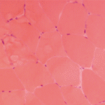
Inflammation involves the endomysium (i.e., around individual muscle fibers in the fascicles), or the muscle fibers themselves. The rimmed vacuoles for which inclusion body myositis is named are best observed with modified Gomori trichome stain. Of note, rimmed vacuoles may also be seen in other neuromuscular disorders.2
Immune-mediated necrotizing myopathy, which is most commonly associated with anti-signal recognition particle (anti-SRP) and anti-3-hydroxy-3-methylglutaryl coenzyme A reductase (anti-HMGCR) antibodies, is characterized by random atrophy (e.g., mixed necrotic and regenerating fibers) and a relative paucity of inflammation.
“Compare this with [dermatomyositis], where the pattern is clearly perifascicular,” Dr. Pytel said.
Inflammatory myopathies have distinct but overlapping patterns, so when morphologic features overlap, pathologists can’t always offer a definitive diagnosis.
Don’t Forget the Drugs
It’s important to remember that medications can also cause muscle injury, and many of these toxicities have specific histologic findings. Example: Chronic steroids cause type 2 muscle fiber atrophy. Hydroxychloroquine causes a vacuolar myopathy with lysosomal aggregates visible with staining and curvilinear bodies visible on electron microscopy.
What’s the Diagnosis?
Inflammatory myopathies have distinct but overlapping patterns, so when morphologic features overlap, pathologists can’t always offer a definitive diagnosis.3
“In cases that aren’t clear-cut, immunohistochemical stains can provide additional information, and we’re discovering more of these each year,” Dr. Pytel said. “That’s why polymyositis is becoming a vanishingly small entity.”4
What about biopsies with mild myopathic changes? “A lot depends on clinical correlation,” Dr. Pytel said. “Is the biopsy representative of what led you to do the biopsy in the first place? A muscle biopsy with mild myopathic changes but no inflammatory infiltrates could be due to patchy distribution of disease and biopsy sampling error. Or perhaps it’s [immune-mediated necrotizing myopathy], in which case the absence of inflammation is expected. I’d also consider metabolic myopathy, dystrophies or toxic-metabolic damage. The clinical history is what provides the answers, so the more info you include with the specimen, the better.”
Summary
Dr. Pytel shared high-yield information that can help us better understand muscle pathology reports. Muscle biopsy may not always yield a definitive diagnosis due to morphologic overlap among myopathies. But when provided with ample clinical context, pathologists can refine their interpretations and provide information that allows us to better treat our patients.
Samantha C. Shapiro, MD, is an academic rheumatologist and an affiliate faculty member of the Dell Medical School at the University of Texas at Austin. She received her training in internal medicine and rheumatology at Johns Hopkins University, Baltimore. She is also a member of the ACR Insurance Subcommittee.


