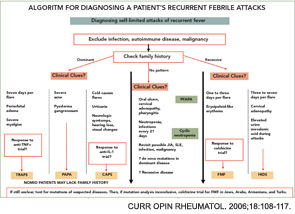
More than 50 mutations in the Mediterranean fever (MEFV) gene have been associated with FMF.2 The MEFV gene consists of 10 exons, and most of the disease-causing mutations are located in exon 10. The gene codes for pyrin, an important protein of the inflammasome and the inflammatory pathway. Pyrin regulates IL-1 production through its interaction with caspase-1; Chae et al have suggested a direct, ASC (apoptosis-associated speck-like protein with a caspase-recruitment domain)-independent effect of pyrin on IL-1β activation and a direct interaction of pyrin with caspase-1 to modulate IL-1β production.7 Thus, mutations in pyrin are associated with increased IL-1β production. Importantly, the IL-1 receptor antagonist anakinra can suppress acute-phase proteins in a patient with FMF and amyloidosis.7
Clinical Features
FMF typically manifests in childhood with recurrent, self-limited attacks of fever in association with abdominal pain, arthritis, or chest pain; these clinical manifestations can occur alone or together.1-3,8,9 The clinical hallmark is an attack in the form of fever and serositis along with elevation in acute phase reactants. The typical attack of FMF, which lasts one-half to three days, is accompanied by fever.1,3,8 Myalgia may occur.9 In the majority of the patients, serositis manifests itself as abdominal pain which can be incapacitating. Arthritis or arthralgia is also very frequent and was present in half of the patients in a large series reported from Israel and Turkey.8,9 Pleurisy in the form of chest pain is common; it is usually unilateral and often spreads to the shoulder.
Rash is not expected in FMF except when it is associated with vasculitis and the erysipelas-like erythema (ELE) which is provoked by pressure-bearing exercise (see Figure 1, p. 20). ELE may not necessarily accompany an attack. In fact, some manifestations of FMF are not associated with attacks. These include protracted febrile myalgia, hematuria, arthralgia, and exertional leg pain.8 Another notable feature of FMF is the association with vasculitis and, especially, polyarteritis nodosa.10 A large series from Turkey has shown that FMF patients have increased rates of Henoch-Schonlein purpura, polyarteritis nodosa, and probably Behçet disease.9
Laboratory Findings
While spontaneous rises and falls of erythrocyte sedimentation rate (ESR), C-reactive protein (CRP), and fibrinogen are quite characteristic of the attacks in FMF, substantial subclinical inflammation occurs widely and over prolonged periods in patients with FMF.10,11 These findings suggest that the relatively infrequent clinically overt attacks represent the tip of the iceberg in this disorder: Lachman et al have shown that there was also inflammatory activity between attacks as well (median serum amyloid A [SAA] 6mg/l, CRP 4 mg/l).11 SAA levels have proven to be the most informative in identifying the level of inflammation. In a study from our center, my colleagues and I have shown that SAA levels are also associated with an insufficient dose of colchicine, and we use SAA levels to monitor disease control.


