The motions of this unique joint can be appreciated through palpation. To accurately locate the thumb CMC joint and feel the kinematics of abduction, palpate the lateral surface of the head of the thumb metacarpal at the metacarpophalangeal (MP) joint. Slide the palpating digit down to the flared base of the thumb metacarpal. Adduct the thumb while maintaining this palpating position. As described above, the base of the metacarpal will glide laterally and become prominent under the palpating digit. The trapezium is just proximal to the base of the metacarpal and remains stationary during the movement. To palpate flexion of the thumb across the palm, move from the lateral position to the base of the thumb metacarpal on the dorsal aspect. The metacarpal base can be palpated as it rolls and glides towards flexion.
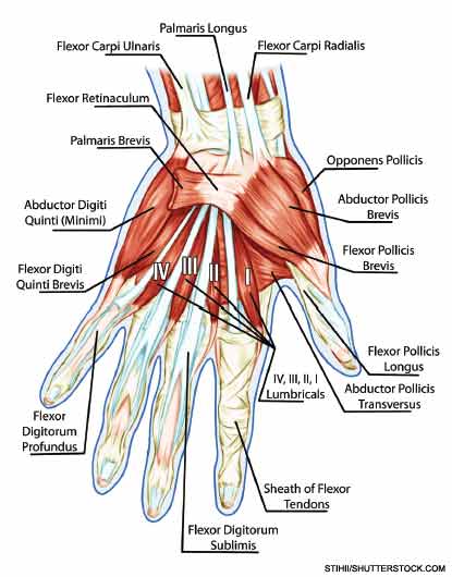
Muscle activity: Muscles that cross the thumb CMC joint include the flexor pollicis longus and brevis, adductor pollicis, abductor pollicis longus and brevis, extensor pollicis longus and brevis, and opponens pollicis (see Figure 1). The median nerve-innervated thenar muscle group is composed of the opponens pollicis, abductor pollicis brevis, and half of the flexor pollicis brevis. The primary purpose of the abductor pollicis longus and brevis, and the extensor pollicis longus and brevis, is to position the thumb. An example would be opening the hand in preparation to grasp a cylindrical glass. These muscles do not typically produce significant forces. The opponens pollicis also positions the thumb, rotating the pad of the thumb to face the pads of the fingers in preparation for pinching activities. The muscles that supply significant force during grasp or pinch activities are the flexor pollicis longus and brevis, and adductor pollicis.

Pathomechanics of the Thumb CMC Joint
Ligamentous laxity and joint impingement: The movements at the thumb CMC joint explain the characteristic problems seen with thumb CMC joint OA. Laxity in the anterior oblique or “beak” ligament can predispose the thumb CMC joint to excessive pressure on the volar portion of the trapezium, particularly during lateral pinch activities such as holding a key.5 With beak ligament laxity, resisted pinching can lever the base of the metacarpal dorsally, resulting in volar compression and posterior translation of the base of the metacarpal. The dorsal/lateral subluxation of the metacarpal base produces the “shoulder sign,” a lateral prominence of the CMC joint, which has been associated with CMC OA (see Figure 2).


