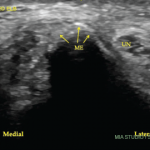
When a meniscus extrusion is symptomatic, patients often report joint line pain that is severe, constant, worse with weight bearing, and when medial, the patient often has to sleep with a pillow between the knees to limit contact. On examination, the affected joint line may appear swollen and the joint line and adjacent femur and tibia are exquisitely tender, particularly along the mid and anterior portion of the joints.
Plain radiographs of a knee with a meniscus extrusion demonstrate narrowing of the joint space with or without osteophytes and flattening of the femoral condyle and squaring and sclerosis of the tibial plateau. On MRI, the extruded meniscus may be displaced superiorly or inferiorly in the medial or lateral gutters. Frequently, MRI shows adjacent edema in the medial collateral ligament (MCL) and its bursa medially (see Figure 1, left). This MCL edema may be due to several factors including the pressure of the extruded meniscus on the local capsule and ligament, inflammation of the synovio-entheseal complex, local friction from osteophytes against the MCL during knee movement, and increased force upon the MCL due to changes in knee mechanics (increased load transmission, loss of shock absorption, and decreased congruency of the femorotibial joint) caused by the meniscal extrusion.4-7 MRI frequently demonstrates bone marrow edema in the femoral condyle and tibial plateau adjacent to the extruded meniscus (see Figure 2B, above). It is worth noting that bone marrow edema is also a risk factor for rapid structural deterioration in knee osteoarthritis; its relation to progression may be explained in part by its association with limb malalignment.8-10
Determining whether a patient has a symptomatic extrusion of the meniscus, rather than arthritis alone, has important implications for treatment. The extruded meniscus may not improve with typical non-operative treatments for arthritis, such as physical therapy and exercise, corticosteroid injections, or other medications. More aggressive treatment can be considered to improve the biomechanical environment in the affected compartment. Unloader braces may help pain by decreasing mechanical stress by reducing malalignment and improving joint stability.11 The use of bisphosphonates to treat bone marrow edema and perhaps to decrease subchondral bone turnover associated with OA can be considered, although this approach is controversial.12
Arthroscopic surgery has a place in managing the arthritic knee with a symptomatic meniscus extrusion. While arthroscopic surgery may be controversial for the treatment of arthritis in general (i.e., lavage alone for relief of pain), surgery remains beneficial for treating mechanical symptoms associated with meniscus tears, delaminating chondral lesions and loose bodies. In the case of a meniscus extrusion, arthroscopic meniscus debridement may provide relief by decreasing impingement of the thickened, extruded meniscus upon local structures such as the MCL (see Figure 2, above).
