Skin Deep
On a different subject, Lisa Zickuhr, MD, MHPE, associate program director of the rheumatology fellowship training program, Washington University, St. Louis, Mo., and Sarah Goglin, MD, associate program director of the rheumatology fellowship training program, University of California, San Francisco, described their work to create a module to teach the assessment of cutaneous lupus erythematosus across differing skin tones. The objectives fellows are meant to achieve via the module are fourfold:
- To identify acute, subacute and chronic cutaneous lupus in a variety of skin tones;
- To correlate the pattern of cutaneous lupus with the differential risk of systemic disease;
- To differentiate acute, subacute and chronic cutaneous lupus from mimicking conditions; and
- To develop strategies that accurately photograph cutaneous lesions in patients with skin of color.
Dr. Zickuhr cited research indicating patients with skin of color are underrepresented with respect to educational photos of cutaneous findings of lupus and related conditions.1,2 Such underrepresentation is potentially associated with decreased clinician confidence in identifying lupus-related rashes in patients with skin of color.3
The module is an online, asynchronous, interactive case-based program designed to be used in programs around the country. A number of different conceptual frameworks were employed in the module’s design. Content layering involves starting simple and building in complexity from that initial foundation. This framework is illustrated in the way the module starts with a pre-test and then incorporates a flowchart that introduces the different forms of cutaneous lupus, which is then followed by a post-test to gauge comprehension. The program employs case-based learning with a simulated patient who presents with subcutaneous lupus and asks a series of questions to the clinician—in this case a trainee taking the module—who must select the most appropriate response to the patient.
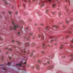
Relying on the power of visual representation, Dr. Zickuhr, Dr. Goglin and colleagues sought to use as little text as possible to describe cutaneous lupus findings and used photographs and drawings to illustrate its classic findings. Additionally, the program has an audiovisual component. When the fellow is shown a histopathologic slide demonstrating interface dermatitis, the slide is accompanied by an audio file that details the salient features of this slide.
Interleaving involves taking similar but different concepts and showing examples side by side so that nuances can be compared. In the module, interleaving is used by showing photos of patient faces with acute cutaneous lupus findings placed next to each another, with variations in skin tone across the photos.


