Highlights from the 2017 EULAR Congress
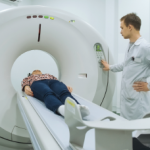
MADRID—Researchers say that whole-body MRI could yield an earlier diagnosis of spondyloarthropathy (SpA) in patients with early inflammatory joint symptoms, according to findings presented in a poster session at the Annual European Congress of Rheumatology (EULAR). The approach could lead to earlier treatment and better outcomes, they say.
Investigators at the University of Leeds recruited 39 patients, an average of 43 years old, with early inflammatory joint symptoms, signs of inflammatory arthritis or both.1
An array of clinical data was collected, and patients were categorized clinically: 14 were deemed to have definite inflammatory arthritis in the form of either rheumatoid arthritis (RA) or SpA, and 25 had undifferentiated arthritis (UA).
They underwent 3.0 Tesla, T2-weighted fat-suppressed MRI of the spine and sacroiliac joints before IV contrast, and 3D VIBE Dixon imaging of the peripheral joints and enthuses postcontrast.
Patients in the CCP-positive RA group were found to have disease activity mainly in the small joints and tendons. Those in the CCP-negative group had disease activity that was large-joint based, with half having sacroiliac joint disease.
In the undifferentiated group, patients with persistent UA had disease distributed in both the axial and peripheral areas, involving both the joints and the enthuses, with a quarter having sacroiliac joint disease. Among those with resolved UA, there was a similar distribution of disease, but with no sacroiliac disease.
A year after the imaging, six more patients were diagnosed with SpA—three patients in the CCP-negative RA group and three in the persistent UA group, all of whom had baseline MRI findings that were similar to the patients already diagnosed with SpA at baseline.
Using whole-body MRI could make a difference for patients, especially those in the antibody-negative group, said Jane Freeston, MD, a consultant rheumatologist at the University of Leeds.
“The options for the group that don’t quite fit nicely into normal patterns [can be limited],” she said. “They do badly because no one can really work out what’s going on. And this offers a better way to understand what’s wrong with them and then treat them earlier and more appropriately.”
Ultrasound Reveals Active Inflammation
In another study, researchers found that RA patients in long-standing clinical remission after being treated with a DAS28 treat-to-target approach still had signs of active inflammation on ultrasound, whether they’d been treated with conventional synthetic disease-modifying anti-rheumatic drugs (csDMARDS) or biological DMARDS.2
The study illustrates again that inflammation is ongoing even in patients with no other sign of the disease, but researchers are waiting on further results to try to tell whether the inflammation is predictive of progression, said Uffe Moller Dohn, MD, PhD, a senior consultant in rheumatology at the Copenhagen Center for Arthritis Research.
Previous research from investigators at the University of Leeds found that inflammation on ultrasound was predictive of progression in RA patients, but the criteria for remission in that study were less strict, and many of the patients would not have qualified as being in remission by DAS28, Dr. Dohn said.3
In the study at his center, 87 RA patients, in continuous DAS28 remission for at least a year and with no radiographic progression for at least a year, had ultrasound imaging on 24 joints. The joints were scored 0 to 3 for grey-scale synovitis (GSS) and synovial color Doppler activity (CDA). Those with scores of 0 on both for all joints were considered to have no inflammation. And those with a GSS score of 1 but still CDA scores of 0 were considered to have minimal inflammation.
Total absence of inflammation on ultrasound wasn’t seen in any of the patients treated with csDMARDS and in just 14% of those treated with biological DMARDS (P=.01). But the treatment groups showed no statistically significant difference in minimal inflammation, seen in 33% and 40%, respectively. A CDA score of at least 1 in at least one joint was seen in more than half of the patients in both groups.
“So even though they were in DAS remission for a long-standing period and even though they did not have radiographic progression [for a year], we still found active inflammation in the majority of patients,” Dr. Dohn said.
He hopes they will eventually be able to tell whether the inflammation findings can help identify the patients at risk.
“We continue to identify them and see whether they progressed,” he said.
Subclinical Inflammation on MRI in PsA
Researchers in a third study are assembling data in a relatively unexplored area—subclinical inflammation on MRI in the feet of patients with psoriasis—in an effort to find predictors of development of psoriatic arthritis (PsA), said Ashish Mathew, MD, assistant professor at Christian Medical College in Vellore, India.4
Fifty-three patients with psoriasis without arthritis (PsO) and 30 patients with PsA were enrolled, and their feet were imaged with a low-field extremity MRI. Researchers found high percentages of patients with subclinical inflammation; it was seen in 64% of the PsO patients and in 67% of the PsA patients, not a statistically significant difference. They also found that a score of greater than 3 on the Early Arthritis in Psoriasis questionnaire was a predictor of subclinical foot inflammation (P=.008).
About 40% of patients with psoriasis end up being diagnosed with psoriatic arthritis, and early treatment leads to better outcomes. Dr. Mathew said his team hopes to be able to draw some significance from the inflammation findings over time.
“Does that mean anything? Not at the moment,” he said. “What we’re essentially doing is following them to see how many of them develop psoriatic arthritis.”
Thomas R. Collins is a freelance writer living in South Florida.
References
- Freeston JE, Mankia KS, D’Agostino MA, et al. Can whole body MRI at baseline identify definite inflammatory arthritis patterns in undifferentiated arthritis? (abstract OP0630). Annual European Congress of Rheumatology. 2017 Jun 16. Madrid, Spain.
- Dohn UM, Brahe CH, Hetland ML, et al. Ultrasound shows signs of inflammation in most patients with rheumatoid arthritis in longstanding clinical remission, irrespective of conventional or synthetic or biologic DMARD therapy (abstract OP0628). Annual European Congress of Rheumatology. 2017 Jun 16. Madrid, Spain.
- Brown AK, Quinn MA, Karim Z, et al. Presence of significant synovitis in rheumatoid arthritis patients with disease-modifying antirheumatic drug-induced clinical remission: Evidence from an imaging study may explain structural progression. Arthritis Rheum. 2006 Dec;54(12):3761–3773.
- Mathew AJ, Bird P, Gupta A, et al. Magnetic resonance imaging (MRI) inflammation of the feet demonstrates subclinical inflammatory disease in cutaneous psoriasis patients without clinical arthritis (abstract OP0629). Annual European Congress of Rheumatology. 2017 Jun 16. Madrid, Spain.

