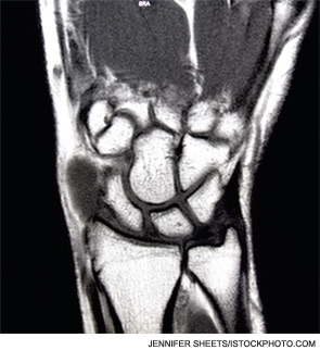
An analysis of wrist MRIs from participants in the Treatment of Early Aggressive Rheumatoid Arthritis (TEAR) clinical trial indicates that patients continue to show joint inflammation even after two years of early aggressive therapy.
Investigators Harold Paulus, MD, and Veena Ranganath, MD, both from the University of California, Los Angeles, initiated the research to determine whether imaging studies could be used to refine the clinical remission criteria for rheumatoid arthritis (RA). At the time, Dr. Paulus was involved in the TEAR study with Larry Moreland, MD, of the University of Pittsburgh. All three investigators received grants from the ACR Research and Education Foundation (REF) Within Our Reach: Finding a Cure for Arthritis program.
“We proposed looking at the patients in the TEAR clinical trial and obtained MRIs of the wrists in patients who completed two years of aggressive treatment with methotrexate plus etanercept, or triple [disease modifying antirheumatic drug] therapy,” Dr. Ranganath says. “This provided us with two years of clinical data from a randomized, controlled clinical trial. We obtained 118 MRIs, and what we found was that despite patients having been on two years of aggressive therapy, we didn’t see a single MRI that was completely devoid of inflammation.” The MRIs revealed synovitis and tenosynovitis.
“A previous observational study of RA patients judged to be in remission by their physicians by Brown and colleagues also reported that residual joint inflammation was present in joint MRIs,” Dr. Paulus says.1 “Our study confirms these findings in predefined, early RA patients who completed a well-controlled, rigorous, two-year clinical trial using aggressive treatment regimens. These findings highlight differences between ‘clinical remission’ and ‘imaging remission.’ Imaging studies can detect persistent signs of synovitis and tenosynovitis in patients with minimal evidence of joint swelling or tenderness.”

Dr. Ranganath says there are a couple of ways to think about this information from a clinical perspective.
“One possibility is that MRIs are too sensitive and we’re picking up on other things unrelated to RA,” she says. “However, this could also mean we shouldn’t be taking our patients off medications until we have better studies that confirm the durability of apparent remission. There certainly may be clinical implications.

I’m not sure we can make definitive recommendations yet, but I think this is an area of research that requires further study.”
Drs. Paulus and Ranganath will continue to analyze data from their work and the TEAR study to shed more light on the relationship between imaging results and RA remission.


