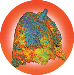
K H FUNG / Science Source
A recent proof-of-concept study to evaluate nuclear imaging in interstitial lung disease (ILD) concludes it is feasible to study ILD subtypes using this technology to visualize specific molecular processes of ILD. The process has important potential applications for the development of targeted molecular therapies.1
ILD is an umbrella term for a group of heterogeneous lung diseases, of which the two most common are idiopathic pulmonary fibrosis and ILD that presents in the context of connective tissue disorders, such as systemic sclerosis. The common end stage of ILD, pulmonary fibrosis, is a chronic, progressive and ultimately fatal lung disease with poor prognosis and high mortality rates.
SPECT Scans
Using molecular biology techniques, the team of researchers, based at University Hospital Zurich and the Center for Radiopharmaceutical Sciences (CRS) in Switzerland, conducted analyses of lung tissue specimens with severely damaged lung architecture obtained through lung transplantation in patients with ILD, along with samples from bleomycin (BLM)-challenged mice and respective controls.2 They performed single photon emission computed tomography (SPECT) scans, a nuclear imaging methodology, to visualize the pulmonary expression of two molecules implicated in the pathophysiology of ILD, integrin alpha-v beta-3 (αvβ3) and somatostatin receptor 2.
Specific pulmonary accumulation of radiotracers was assessed by in vivo and ex vivo scans at several time intervals in ILD. Expression of the two molecules was found to be substantially increased in patients with ILD regardless of subtype, as well as in the BLM-treated mice, with stage-specific overexpression by rates three- to fourfold over controls.
Nuclear imaging produces images from radiation following the introduction of a small amount of a radioactive tracer, typically by injection into a vein. The radiotracer travels through the area being examined and emits energy in the form of gamma rays, which are used to create images of the diseased site in the body. SPECT and positron emission tomography scans are the most common nuclear imaging modalities used in routine diagnostics. These procedures are noninvasive and typically painless medical aids in the diagnosis and evaluation of various medical conditions.
Nuclear imaging has the advantage of truly capturing and visualizing pathophysiology compared with high-resolution computed tomography and magnetic resonance imaging, which reflect anatomy, says the new study’s corresponding author, Britta Maurer, MD, senior consultant rheumatologist and group leader at the Center of Experimental Rheumatology at University Hospital Zurich. She also holds the Prof. Max Cloetta Research Position at the center. Nuclear imaging provides unique information that often cannot be obtained using other imaging procedures and offers the potential to identify molecular disease subtypes.
