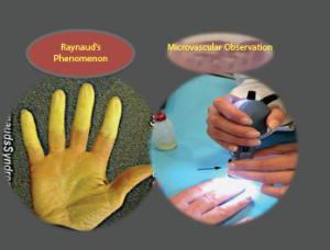
The connective tissue disease most frequently associated with Raynaud’s phenomenon is systemic sclerosis, followed by mixed connective tissue disease, but it’s also sometimes seen in lupus, rheumatoid arthritis, Sjögren’s syndrome and others. It is also important for clinicians to recognize that Raynaud’s phenomenon sometimes accompanies other disease conditions, such as vasculitis, hematological disorders and malignancy.7
It’s extremely important to differentiate patients with primary Raynaud’s phenomenon from those with secondary Raynaud’s phenomenon. Dr. Herrick explains, “Capillaroscopy can allow early diagnosis of systemic sclerosis, because Raynaud’s phenomenon is the most common presenting feature of systemic sclerosis. Normal nailfold capillaries are reassuring. Conversely, definitely abnormal capillaries point to an underlying systemic sclerosis-spectrum disorder. Sometimes the test is equivocal, and sometimes the capillaries cannot be easily seen. If diagnostic uncertainty remains, then consideration should be given to repeating the capillaroscopy in six to 12 months.”

Raynaud’s syndrome.
Along with capillaroscopy, clinical history and exam, clinicians should run basic ANA and ESR tests to rule out the possibility of secondary Raynaud’s phenomenon. If any clinical suspicion remains, physicians should request additional laboratory assessments for connective tissue disease.7
The problem for the clinician is that some patients presenting with primary Raynaud’s phenomenon will eventually transition to secondary Raynaud’s phenomenon. A meta-analysis indicated 20% of patients initially diagnosed with primary Raynaud’s phenomenon later transitioned to secondary Raynaud’s phenomenon over a follow-up period of 10 years.8
Maurizio Cutolo, MD, is professor of rheumatology and director of the research laboratories and clinical academic unit of rheumatology at the University Medical School of Genova, Italy. Dr. Cutolo advocates nailfold viewing of these primary Raynaud’s phenomenon patients every six months. “The nailfold videocapillaroscopic analysis of patients with primary Raynaud’s phenomenon is the most practical, cheap and reliable system to very early detect the development of a connective tissue disease like systemic sclerosis, even before the appearance of specific autoantibodies in the serum.” Other clinicians have recommended such assessments every 12–24 months in patients with primary Raynaud’s phenomenon.2
Patients who have symptoms of primary Raynaud’s phenomenon and who also have systemic sclerosis specific changes on capillaroscopy are very likely to transition to definitive systemic sclerosis (SSc). SSc-specific antibodies (anti-centromere protein B, anti-topoisomerase I, anti-Th/To, or anti-RNA polymerase III) can also be helpful in establishing prognosis. Patients who do not have any SSc-specific autoantibodies or abnormal nailfold capillaroscopy findings are extremely unlikely to ever develop secondary Raynaud’s phenomenon. In contrast, one study found that 80% of patients with both abnormalities developed systemic sclerosis over 20 years of follow-up.9

