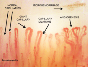
Clinicians can also utilize prognostic indices based on capillaroscopy findings to assess the probability of progression to SSc. Using the Prognostic Index for Nailfold Capillaroscopic Examination (PRINCE), clinicians can use specific findings to estimate the five-year probability of secondary Raynaud’s phenomenon developing in patients with isolated Raynaud’s phenomenon. The presence of giant capillary loops, microhemorrhages and reduced capillary number seem to be the characteristics with the most predictive role.10
Systemic Sclerosis
Identifying patients in the very beginning stages of SSc is important, because early use of vascular remodeling drugs may alter the disease course.8 Currently, no treatment for SSc has been clinically proven to halt the disease progression in people with clinically recognizable disease, but available treatments may make a difference if started earlier, before irreversible damage has occurred.3
In 2000, Dr. Cutolo and colleagues first categorized the morphological patterns that can be observed in SSc.11 These can be used to help stage the disease and evaluate drug therapy. It’s important to note the extent of microvascular damage correlates with the severity of skin, heart and lung involvement.12
Dr. Cutolo notes, “Distinct morphological patterns on nailfold videocapillaroscopy and a gradual increase in severity of microvascular abnormalities (quantifiable and scored) are observed during clinical progression of systemic sclerosis and seem to reflect the evolution of the disease process.” The changes reflect the pathophysiological changes characteristic of the disease, with hypoxia over time leading to capillary modification and neo-angiogenesis directed by growth factors in a profibrotic environment.5
An early pattern is characterized by the appearance of several dilated capillaries, some bleeding, but without loss of capillary loops. This early pattern can be often be detected many years before the full clinical manifestations of SSc become apparent.2
In somewhat more advanced disease, an active pattern appears, displaying frequent bleeding megacapillaries, moderate loss of capillaries, moderate disorganization of the capillary architecture and rare or absent branching capillaries.3
Even more advanced scleroderma is characterized by a late pattern, which shows severe capillary loss, neovascularization by megacapillaries and massive avascular areas. This late pattern is more frequent in patients who have active disease and moderate to severe skin or visceral involvement.11
In general practice, clinicians generally categorize these patterns qualitatively based on pattern recognition. However, there are semi-quantitative methods and quantitative methods that can be used to assess certain characteristics of the capillaries using manual or automatic methods. Researchers have also used scoring systems to assess the risk of digital ulceration as a complication. Other studies have looked at the association between capillaroscopic findings and the onset of interstitial lung disease, pulmonary artery hypertension, cardiac involvement, skin involvement and death.3,12
Pathogenic changes in capillary morphology may long predate the onset of clinical symptoms. And in patients already diagnosed with a systemic disease, such as systemic sclerosis, the level of capillary damage may reflect internal organ involvement.
Evaluation of Treatment Response

Microhemorrhages represent the death of the giant capillaries. Their disappearance from the microvessel array, with consequent loss of capillaries, is followed in advanced stages by angiogenesis and the formation of new, abnormal vessels.

