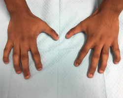
Figure 1. Bilaterally, the hands demonstrate hypoplasia of distal phalanges, hypoplastic nails and flexion of distal interphalangeal joints.
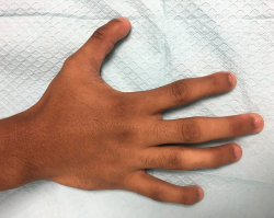
Figure 2. This image of the right hand gives us a closer view of the fourth and fifth digits; however, the mild extension of the proximal interphalangeal joints is not well visualized in the picture.
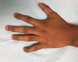
Figure 3. This image of the left hand gives us a closer view of the fourth and fifth digits; however, the mild extension of the proximal interphalangeal joints is not well visualized in the picture.
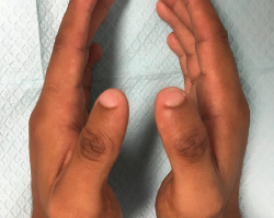
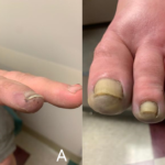
Figure 4. Bilaterally, the thumbs have a thinner-than-normal appearance, with hypoplastic nails.
His feet show hypoplastic distal phalanges and toenails, bilaterally (see Figure 5). He is also found to have flexion deformities of distal interphalangeal joints in his feet (see Figure 6).
X-rays performed in the office reveal hypoplasia of the distal phalanges in his hands (see Figures 7 and 8) and feet (see Figures 9 and 10).
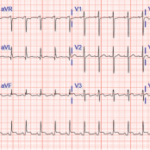
His comprehensive metabolic panel and vitamin D and parathyroid hormone levels are all normal. A review of his transthoracic echocardiogram report from four years earlier is normal, except for trace tricuspid valve regurgitation.
His hand and foot abnormalities are thought to be congenital malformations, and they are also suggestive of fetal hydantoin syndrome. After reviewing many studies supporting this diagnosis and having detailed discussions with the patient and his father, we did not pursue any further workup, including genetic testing. The patient did not have evidence suggestive of any other underlying abnormalities.
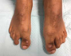
Figure 5. Bilaterally, the feet have hypoplasia of the distal phalange
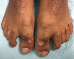
Figure 6. Hypoplastic nails and flexion of the distal phalangeal joints.
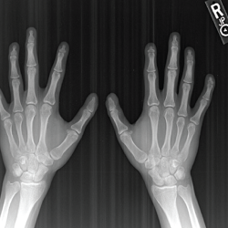
Figure 7. An X-ray of both hands—antero-posterior view—shows hypoplasia of the distal phalanges, which is more pronounced over the second and fifth digits.
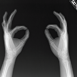
Figure 8. An X-ray of both hands—oblique view—shows hypoplasia of the distal phalanges.

Figure 9. An X-ray of both feet—antero-posterior view—shows hypoplasia of the distal phalanges and subluxation of the distal interphalangeal joints.
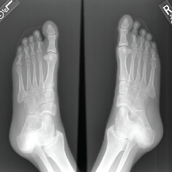
Figure 10. An X-ray of both feet—oblique view—shows hypoplasia of the distal phalanges.