
Before there was MTV, there was Ed Sullivan. His Sunday night variety show hosted some of the finest music and comedy acts that were seen on television. Sullivan had a knack for spotting exceptional talent. His show debuted some of the greatest rock and roll bands of the 1960s, including The Rolling Stones, The Doors, The Who and most famously, The Beatles. Viewed by an audience estimated to be more than 76 million people, about 40% of the U.S. population, the appearance of The Beatles launched the first tidal wave of Beatlemania across America and around the world.
Unlike his musical guests, Sullivan displayed little stage presence, often bungling his lines as he sauntered stiffly across the stage, arms firmly folded across his chest. Comedians would often poke fun at Sullivan’s wooden style, imitating his erect posture and his odd way of turning his entire body rather than just his neck when looking sideways. Many years later, I discovered the reason for Sullivan’s rigid demeanor. He suffered from ankylosing spondylitis (AS), an ancient disease that has only recently attracted the intense interest of clinicians and researchers.
In Search of a Remedy
For patients such as Ed Sullivan, what treatment choices were available that would reduce their insufferable stiffness? Sadly, there were very few. High-dose salicylate therapy, an effective remedy for relieving the achiness of patients with rheumatoid arthritis (RA) was ineffective in the AS population. Similarly, the recently synthesized corticosteroids, so beneficial in treating RA and lupus, provided meager benefits. In the 1960s, the mainstay of AS therapy was phenylbutazone (PBZ), or the bute, the first commercially developed nonsteroidal antiinflammatory drug (NSAID) after aspirin.
Among veterinarians, the bute was a hallowed drug that seemed to cure horse lameness. The bute achieved notoriety in 1968 when Dancer’s Image, the winner of the Kentucky Derby, was disqualified after traces of it, a banned substance during races, were discovered in the horse’s postrace urine test. Dancer’s Image was retired at the age of 4 and spent his remaining years siring many swift thoroughbreds. Unfortunately, along with power and speed, his offspring carried his trait for developing chronically inflamed ankles.
Although many patients considered the bute their panacea for alleviating stiffness and misery, it carried a distressing list of adverse effects, including agranulocytosis and fulminant hepatic necrosis. Not surprisingly, the U.S. Food and Drug Administration banned its use following the release of the next NSAID, indomethacin.
An Unanticipated Consequence of War
The bute wasn’t the only hazardous therapy used to treat AS. The creation of large armies to fight World War II required a massive conscription effort on both sides of the Atlantic. Suddenly, millions of young men were drafted for military duty. As part of the enlistment process, they underwent medical exams that focused on their suitability for duty. For some of these recruits, it was their first encounter with their nations’ healthcare system. Given the huge numbers of draftees, it was not surprising to observe that AS gained prominence as a major rheumatologic disease. In one study, nearly one-fifth of patients with chronic back pain who were admitted to a U.S. Army general hospital were diagnosed with AS.1 But rheumatologists were split regarding the proper clinical designation for AS.
In the U.S., AS was viewed as a variant of RA on the basis of a shared finding of an elevated erythrocyte sedimentation rate and the occasionally damaged peripheral joint. European clinicians argued for the distinction of AS from RA, because of its contrasting disease demographics, such as male preponderance and an earlier age of onset. In addition, the absence of subcutaneous nodules and the lack of response to gold salt therapy favored a unique diagnostic distinction. Ultimately, the discovery of rheumatoid factor and its widespread clinical application confirmed the European opinion.2
Regardless of whether one was a lumper or a splitter, a popular treatment for refractory AS was radiation therapy. The rationale for its use was best summarized by one of its major British advocates:3
There is no need to stress the tremendous economic importance of satisfactory treatment of this disease attacking as it does young men and women yet to reach their prime. The response to comprehensive radiotherapy is most encouraging and at times even dramatic. Probably it is correct to state that the results prove more gratifying than in any of the benign conditions which the radiotherapist is called upon to treat.
Sadly, it took many years and countless cases of leukemia before radiation therapy was abandoned as a treatment for AS.
The Great Wasteland
The spine is a great wasteland. After all, this sizable chunk of body real estate does not readily lend itself to a careful, critical exam. For many doctors, it fails to attain the prominence of the heart, with its complex array of murmurs, rubs, clicking sounds and palpable heaves. Palpation of the spine fails to excite the examiner the way an enlarged spleen or liver does.
Who really bothers to carefully examine the back? Last week, I had the opportunity to teach our medical students the finer points of the musculoskeletal exam. Although they were familiar with some common sense approaches to the examination of the knee or the shoulder, they were lost when we moved to the back. Yes, one can palpate the spine and marvel at its flexibility or lack thereof, but these observations often provide us with little relevant information. Is there any utility palpating the sacroiliac joints? After all, in patients with back pain, everything seems to hurt down there!
Although the students were intrigued when I pulled out my tape measure to perform the Schober’s test, they were dismayed to learn that its namesake, Paul Schober, served as a physician at a German prisoner of war camp during World War II. We ended the session by demonstrating the arcane exercise of measuring the distance between the occiput of a patient’s head and an adjacent wall. This is where Home Depot meets rheumatology. Is there a more esoteric test in medicine than the occiput-to-wall measurement?
Dawning of a New Era: HLA- B27
Our wonderful colleague, Muhammad Asim Khan, MD, professor of medicine at Case Western University in Cleveland, Ohio, embodies the best of rheumatology. A thoughtful, articulate clinician and a patient himself, he has written extensively on the topic of AS.4 Khan vividly recalls how the link was made between HLA-B27 and AS.5
It starts with a Norwegian immunologist, Erik Thorsby, immunizing a colleague by transplanting skin and transferring cells from a healthy HLA identical donor. He assumed that this donor might possess a “hitherto-undetected HLA antigen.” In fact, his colleague, the recipient of the graft and known by his initials, FJH, produced an antibody that detected the new antigen, which Thorsby named, TH-FJH.
Apparently, Thorsby was not shy about using friends and families in his experiments. He subsequently tested his own family and noticed that one member who carried this new antigen had AS, while another possibly had this diagnosis as well.6 However, because most of his family members who were positive for this new antigen were completely healthy, Thorsby discarded this finding, assuming that it was caused by chance. I suspect that Thorsby’s family and friends were grateful that he reached this erroneous conclusion and abandoned his research before he could wreak havoc with their health.
A few years later, Derek Brewerton, MD, and colleagues at the Westminster Hospital in London, England, proposed to tissue-type patients with AS. Dr. Brewerton was intrigued by the clinical observation that AS could have a genetic predisposition. He was deeply influenced by the work of another colleague, Philip Gofton, MD, who described a strikingly high incidence of AS among the Haida Indians of the Queen Charlotte Islands in British Columbia, Canada.7
Dr. Brewerton reflected how this monumental study got started. “It was lunch in the common room at Westminster Hospital on a hot summer day in 1971. Over the salad we decided to investigate the frequency of HLA antigens in ankylosing spondylitis.”8 Although his proposal was initially turned down by his cash-strapped hospital, he eventually tested eight patients with AS for the HLA-B27 gene and found it present in all of them, a chance of less than one in a million. Bingo!
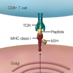
Following the discovery of this critical genetic association, which still represents the strongest link with any complex disease, there was a flurry of research attempting to explain the vital role of the B-27 antigen in the development of AS. Some investigators believed the gene provided some form of molecular mimicry, thus generating an aberrant the immune response. Others speculated a role for the gene in presenting arthritogenic peptides that could have been derived from enteric bacteria. More recently, the focus has turned to the strong genetic association between endoplasmic reticulum aminopeptidase 1 (ERAP1) with HLA-B27-positive AS.
ERAP1 is involved in trimming peptides to optimal length for binding to HLA class 1 molecules, thereby not only affecting the stability and processing of HLA-B27 but also influencing the peptide repertoire presented to the immune system. It appears likely that the pathogenic effect of ERAP1 may be mediated through effects on innate immunity, such as altering the interaction between HLA-B27 and immune receptors or via the endoplasmic reticulum unfolded protein response.9
Hiding in Plain Sight
For many patients and their clinicians, making the diagnosis of AS can remain elusive. The clues are not necessarily found at the bedside. Instead, they may be lurking elsewhere, hiding somewhere in the patient’s history. Before we look, we must listen. Is the pain worse with rest and better with activity? Was there a robust response to NSAIDs? Is there a family history of a spondyloarthritis?
The true number of patients with AS and related forms of inflammatory back disease is hard to ascertain. One recent study from the Netherlands estimated that nearly one-quarter of relatively young patients with chronic back pain seen in primary care practices suffer from an undiagnosed spondyloarthritis.10
I recently met a patient who was referred to me for further management of his recently diagnosed AS. Phil, a recently retired Wall Street financier, kept active by skiing in Wyoming and fox hunting in Delaware. After falling off his horse during a particularly intense fox chase, his world nearly came to a crashing end. It took a neurosurgeon six hours to meticulously repair Phil’s three fractured cervical vertebrae and stanch the profuse bleeding. He told Phil he was a “tough case” because of his AS. What?
Phil was baffled. He had never heard of AS before. Yes, he realized that he walked stiffly, “sort of like Ed Sullivan,” but he attributed this to having tight muscles. None of his doctors contradicted this theory, although Phil couldn’t remember ever having his back carefully examined. When I asked him, he recalled suffering through two prior episodes of uveitis. But neither his ophthalmologist nor his Upper East Side (N.Y.) concierge internist tied all of these can’t miss clues together. They just lay there, buried in his medical records; disparate bits of clinical information waiting for someone in an operating room to lasso together.
Simon M. Helfgott, MD, is associate professor of medicine in the division of rheumatology, immunology and allergy at Harvard Medical School in Boston.
Timed Up & Go Test for Predicting Falls in the Elderly
Clinical question: How reliable is the Timed Up and Go Test in predicting falls in the elderly?
Bottom line: The Timed Up and Go Test (TUG) is not very reliable at predicting falls in the elderly. However, clinicians can probably gain insight from observing how an older individual performs the task. LOE=1a, systematic reviews (with homogeneity) of randomized controlled trials.
Reference: Schoene D, Wu SM, Mikolaizak AS, et al. Discriminative ability and predictive validity of the timed up and go test in identifying older people who fall: Systematic review and meta-analysis. J Am Geriatr Soc. 2013;61(2):202–208.
Study design: Meta-analysis (other)
Funding source: Unknown/not stated
Setting: Outpatient (any)
Synopsis: In the U.S., physicians seeing elderly patients for a “Welcome to Medicare” visit are required to perform a fall risk assessment. The TUG is the most commonly recommended screening tool for this, even though it was originally developed to measure functional mobility and not to predict falls. To perform the TUG, the physician times, in seconds, how long it takes for an elder to arise from a chair with armrests, walk three meters with any usual assistive devices, turn, return to the chair and sit down. Essentially, the TUG indirectly measures strength, balance and gait stability. However, it does not reflect poor vision, cognition and medication effects, all of which are common causes of falls. The authors systematically searched multiple databases to identify studies that compared the TUG performance in elders who fall and those who don’t. Two authors independently evaluated studies for inclusion and extracted data, and resolved disagreements with a third member of the team. Although they describe their criteria for assessing the quality of studies, the paper does not report the steps taken to assign the quality ratings. Finally, they don’t report looking for unpublished studies and did not assess the potential for publication bias. Ultimately, they included 53 studies with between 12 and 1,200 patients (N=12,832) with follow-up between four weeks and five years. The studies included comparisons of various combinations of fallers, nonfallers and multiple fallers. Overall, the diagnostic accuracy of TUG in classifying fallers and nonfallers was poor. Additionally, when factoring in other things that might be associated with falls, the TUG does not predict future falls. In high-functioning patients, the average TUG time was approximately 10 seconds and the difference in TUG times for fallers and nonfallers was only approximately half a second. In institutionalized elders, the average TUG time ranged between 15 and 52 seconds for fallers, and 12 and 30 seconds for nonfallers. The average difference was approximately 3.5 seconds, which is not likely to help the clinician risk-stratify patients. To underscore this, the authors report the diagnostic accuracy of TUG times: The TUG time was not much better than flipping coins.
Henry C. Barry, MD, MS, professor, Michigan State University, East Lansing.
Copyright© 2014 John Wiley & Sons, Inc.
References
- Boland EW, Present AJ. Rheumatoid spondylitis. A study of one hundred cases, with special reference to diagnostic criteria. JAMA. 1945;129:843–846.
- Spencer DG, Sturrock RD, Buchanan WW. Ankylosing spondylitis: Yesterday and today. Medical History. 1980;24:60–69.
- Richmond JJ. The importance of radiotherapy in the treatment of ankylosing spondylitis. Proc Roy Soc Med. 1951;44:443–447.
- More than a doctor. A life guide to ankylosing spondylitis. Video posted Feb. 4, 2014. http://www.everydayhealth.com/health-report/sanjay-gupta/a-life-guide-to-ankylosing-spondylitis.aspx.
- Khan MA. Remarkable polymorphism of HLA-B27: An ongoing saga. Curr Rheumatol Rep. 2010;12:337–341.
- Thorsby E. HL-A antigens and genes. A study of unrelated Norwegians. Vox Sang. 1969;17:81–92.
- Gofton JP, Robinson HS, Price GE. A study of rheumatic disease in a Canadian Indian population. Ann Rheum Dis. 1964;23:364–371.
- Brewerton, DA. Discovery: HLA and disease. Curr Opin Rheum. 2003;15: 369–373.
- Cheidel S, Chen L, Pointon J, et al. ERAP1 and ankylosing spondylitis. Curr Opin Immunol. 2013;25:97–102.
- van Hoeven L, Luime J, Han H, et al. Identifying axial spondyloarthritis in Dutch primary care patients, ages 20–45 years with chronic low back pain. Arthritis Care Res. 2014;66:446–453.


