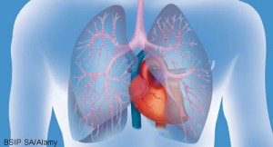
 Sarcoidosis, sometimes referred to as the great mimicker, presents in myriad ways. At the 2021 ACR State-of-the-Art Clinical Symposium, Marc Judson, MD, chief, Division of Pulmonary and Critical Care Medicine, Albany Medical College, New York, provided an update on the screening and treatment of sarcoidosis in patients. He also discussed drug-induced sarcoidosis-like reaction (DISR), a condition he said should be in the deferential for patients in whom sarcoidosis is suspected.
Screening
Dr. Judson noted that the heart, eyes and vitamin D regulation pathway are important places to begin screening for sarcoidosis.
The heart: Among patients with known extra-cardiac sarcoidosis, such symptoms as palpitations lasting more than two weeks, presyncope or syncope, and signs of heart failure should prompt an evaluation for potential cardiac involvement. Objectively abnormal findings may be present on an echocardiogram (e.g., depressed ejection fraction, wall motion abnormalities) or on an electrocardiogram (e.g., first, second- or third-degree atrioventricular nodal block, bundle branch block or supraventricular arrhythmias).
Although proposed screening algorithms differ by organization, Dr. Judson presented evidence that cardiac magnetic resonance imaging (MRI) may be particularly useful. In a study of more than 300 patients with biopsy-proven sarcoidosis, cardiac MRI was found to have higher sensitivity and specificity for the detection of cardiac sarcoidosis than conventional cardiac testing with echocardiogram and electrocardiogram (ECG).1 Detection of cardiac sarcoidosis is of particular importance, because—in contrast to the slow progression of pulmonary fibrosis that can occur in patients with sarcoidosis over months to years—cardiac involvement in sarcoidosis can lead to fatal arrhythmias and sudden cardiac death.
Eyes: With regard to ophthalmic involvement in patients with sarcoidosis, the rationale for screening for sarcoidosis-related eye disease in asymptomatic patients is that this common manifestation of the disease can result in serious consequences even without warning signs.
In a study of 50 patients with biopsy-proven lung sarcoidosis, ocular findings included episcleritis, iris nodules, cataracts, periphlebitis, periarteritis, epiretinal membrane and branch retinal vein occlusion—and more than one in 10 patients lacked ocular symptoms.2 When signs or symptoms do occur, they may include foreign body sensation, blurred vision, floaters or conjunctival hyperemia.
The most serious & permanent damage related to sarcoidosis is from fibrosis. Anti-fibrotic agents may mitigate the effects of fibrotic disease in these patients.
Vitamin D: Vitamin D dysregulation can occur in sarcoidosis. Although calcitriol, the metabolically active form of vitamin D, is normally synthesized entirely in the kidneys, an extra-renal source of calcitriol in sarcoidosis is the alveolar macrophage. These macrophages contain the 1-α hydroxylase enzyme that converts 25(OH) vitamin D to 1,25(OH) vitamin D (i.e., calcitriol). Thus, patients with hypercalcemia can have low 25(OH) vitamin D, low parathyroid hormone and normal to high 1,25(OH) vitamin D.
In addition to hypercalcemia, patients with sarcoidosis can present with hypercalciuria and nephrolithiasis. The astute clinician who notices and evaluates these patterns is likely to identify sarcoidosis in patients with otherwise unexplained hypercalcemia, nephrolithiasis or renal insufficiency.
Treatment
Treatment of sarcoidosis represents an interesting clinical conundrum. The presence of sarcoidosis does not necessarily mandate treatment because the disease does not cause symptoms or organ dysfunction in all patients, and treatments, such as corticosteroids, may have significant adverse effects. Moreover, sarcoid granulomas are believed to result from the interaction of an antigen with the immune system, and granulomatous inflammation may be an attempt to clear the antigen.
Thus, when medications used to treat granulomas are later withdrawn, the inciting antigen may still be present and could lead to granulomatous inflammation and relapse of disease. This process may explain why relapses appear more commonly in patients previously treated for sarcoidosis than in patients who were not previously treated and experienced spontaneous resolution of their symptoms.3,4
When there is physiologic impact of disease, such as shown on pulmonary function testing, or functional impairment due to symptoms and decreased quality of life, Dr. Judson indicated treatment is warranted.
Corticosteroids remain the mainstay of sarcoidosis treatment, but other medications that show some evidence of efficacy include methotrexate, azathioprine, leflunomide, hydroxychloroquine, infliximab and adalimumab.5
Although much of the focus on the treatment of sarcoidosis is on active inflammation, it should be noted the most serious and permanent damage related to sarcoidosis is from fibrosis. Anti-fibrotic agents may mitigate the effects of fibrotic disease in patients with sarcoidosis.
In a pivotal clinical trial of nintedanib for progressive fibrosing interstitial lung diseases, patients receiving this medication had an annual rate of decline in forced vital capacity that was significantly lower than that experienced by the placebo control group. In this trial, about 12% of the study patients had sarcoidosis.6
Given these data, anti-fibrotic agents may be a worthwhile consideration for some patients with advanced sarcoidosis, according to Dr. Judson. But the ongoing development of potential biomarkers is needed to screen for and predict the development of fibrotic disease in patients with sarcoidosis.
DISR
Dr. Judson concluded the talk with a discussion of DISR, which involves a systemic granulomatous reaction that is indistinguishable from sarcoidosis and occurs in a temporal relationship to an offending drug. DISRs can manifest with many of the exact same features of sarcoidosis, including bilateral hilar adenopathy, uveitis, granulomas in scars and tattoos, hypercalcemia and elevated serum angiotensin-converting enzyme (ACE) levels. Additionally, the histology of biopsy samples from these patients is indistinguishable from sarcoidosis.
DISRs may resolve after discontinuation of the offending agent and recur if the agent is reintroduced. Among the medications that more commonly cause DISR are immune checkpoint inhibitors, highly active anti-retroviral therapies, tumor necrosis factor- α antagonists and BRAF inhibitors. It’s important to keep drug reactions in mind when evaluating patients with sarcoid-like symptoms and signs.
In Sum
A great deal remains to be discovered about sarcoidosis. For rheumatologists and other clinicians, this disease will continue to be part of the deferential for many patients, and hopefully, the coming years will allow for its effective diagnosis and treatment.
Jason Liebowitz, MD, completed his fellowship in rheumatology at Johns Hopkins University, Baltimore, where he also earned his medical degree. He is currently in practice with Skylands Medical Group, N.J.
References
- Kouranos V, Tzelepis GE, Rapti A, et al. Complementary role of CMR to conventional screening in the diagnosis and prognosis of cardiac sarcoidosis. JACC Cardiovasc Imaging. 2017 Dec;10(12):1437–1447.
- Pefkianaki M, Androudi S, Praidou A, et al. Ocular disease awareness and pattern of ocular manifestation in patients with biopsy-proven lung sarcoidosis. J Ophthalmic Inflamm Infect. 2011 Dec;1(4):141–145.
- Gottlieb JE, Israel HL, Steiner RM, et al. Outcome in sarcoidosis. The relationship of relapse to corticosteroid therapy. Chest. 1997 Mar;111(3):623–631.
- Rizzato G, Montemurro L, Colombo P. The late follow-up of chronic sarcoid patients previously treated with corticosteroids. Sarcoidosis Vasc Diffuse Lung Dis. 1998 Mar;15(1):52–58.
- Judson MA. Advances in the diagnosis and treatment of sarcoidosis. F1000Prime Rep. 2014 Oct 1;6:89.
- Flaherty KR, Wells AU, Cottin V, et al. Nintedanib in progressive fibrosing interstitial lung diseases. N Engl J Med. 2019 Oct 31;381(18):1718–1727. Epub 2019 Sep 29.
