BALTIMORE—At the 20th Annual Advances in the Diagnosis and Treatment of the Rheumatic Diseases symposium at Johns Hopkins School of Medicine, Jun Kang, MD, assistant professor of dermatology, Johns Hopkins School of Medicine, Baltimore, regaled the audience with his talk, Dermatology and Rheumatic Disease. He discussed a stepwise approach to evaluating skin lesions and helping patients receive proper diagnoses and treatments.
Assessment
Dr. Kang began by discussing primary lesion morphology analysis, a method for describing and categorizing skin lesions based on their shape, size, color and other characteristics. Dermatologists routinely employ this technique and rely on heuristics to quickly arrive at a diagnosis. However, for rheumatic diseases, such shortcuts may be less reliable. A clinician is more likely to arrive at the correct diagnosis if they have seen the presentation before and if they understand how to correlate the histologic findings on skin biopsy with the patient’s clinical course.
Dr. Kang noted that patients may require multiple skin biopsies for diagnosis because inflammatory skin diseases can have overlapping clinical and histologic findings, with nuanced variations. The timing and location of biopsy may also influence the interpretation of the results. Although clinicians generally believe in searching for a unifying diagnosis, one must be aware of Hickam’s dictum, which states that patients “can have as many diseases as they damn well please.”1
CLE
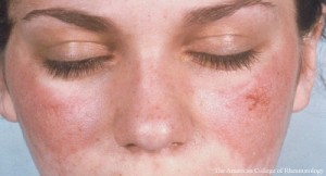
Cutaneous lupus erythematosus (CLE) may be: 1) acute, commonly presenting with a malar rash; 2) subacute, which often manifests with a scaly, red, raised, non-scarring rash in sun-exposed areas; or 3) chronic, such as discoid lupus, lupus panniculitis or chilblain lupus.
The majority of patients with systemic lupus erythematosus (SLE) (70–85%) have skin involvement over the course of their disease. Skin involvement is the presenting sign of disease in 25% of cases.2
In Dr. Kang’s view, essentially all patients with acute CLE have SLE. He explained that skin lesions can sometimes be challenging to recognize in lupus because early scalp discoid lupus erythematosus (DLE) can mimic the pre-scarring phase of alopecia areata and the histologic progression of chronic CLE can mimic morphea. The detection of interface dermatitis on biopsy can be helpful because nearly all forms of CLE are unified by this finding, with the exceptions of tumid lupus and lupus profundus/panniculitis. However, interface dermatitis is not unique to lupus and can be seen in dermatomyositis, Stevens-Johnson syndrome (SJS), toxic epidermal necrolysis (TEN), erythema multiforme, graft-vs.-host disease, mycosis fungoides and other conditions.
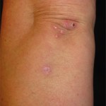
Dr. Kang noted that clinicians must be aware of unusual presentations and complications of CLE. He shared the story of a patient whose clinical presentation of acute CLE mimicked that of SJS/TEN, a condition in which patients may experience wide areas of sheet-like skin and mucosal loss. To distinguish between the two diseases, Dr. Kang noted that SJS/TEN-like acute CLE tends to have a slower progression, less mucosal involvement, more significant alopecia and less full epidermal necrosis on histology than SJS/TEN.
Another entity that clinicians may be unaware of is subacute panniculitis‐like T cell lymphoma (SPTCL), a rare cytotoxic primary cutaneous lymphoma that can be challenging to differentiate from lupus erythematosus panniculitis and may occur in patients with SLE. Although indolent cases of SPTCL can be treated with agents, such as systemic corticosteroids, methotrexate, retinoids, cyclosporine and mycophenolate mofetil. Chemotherapy is indicated for more aggressive disease that behaves like gamma–delta T cell lymphoma.3
Treatment
The treatment paradigm that Dr. Kang employs for CLE begins with photoprotection, smoking cessation and topical therapies. The next step in treatment is anti-malarials, followed by disease-modifying anti-rheumatic drugs, such as methotrexate, mycophenolate mofetil and azathioprine.
If patients continue to experience active disease, then lenalidomide or thalidomide can be prescribed. Thereafter, the treatment options include belimumab, tacrolimus or intravenous immunoglobulin (IVIG).
Additionally, evidence exists to support the use of anifrolumab—a human monoclonal antibody to type I interferon receptor subunit 1 that is approved by the U.S. Food & Drug Administration for the treatment of lupus—for the treatment of cutaneous disease. In a study, Chasset et al. enrolled 11 patients with SLE with biopsy-proved, active CLE who had failed at least three currently available medications. These patients were treated with 300 mg of intravenous anifrolumab every four weeks. The study’s primary outcome of CLASI-A 50 (i.e., proportion of partial response at week 16, defined by a decrease of CLE Disease Area and Severity Index activity of at least 50%) was achieved in all patients. A complete response was observed in six of the 11 patients.4
Based on this and other data, Dr. Kang will consider the use of anifrolumab, early—immediately following the failure of anti-malarial therapy in certain patients.
Dermatomyositis
Dr. Kang stated that differentiation of dermatomyositis from CLE depends wholly on clinical context. Doctors must pay attention to the location and distribution of rash and recognize that it is possible for the two conditions to co-occur in the same patient.
Cutaneous dermatomyositis typically demonstrates a milder interface dermatitis than CLE, and a sizable number of patients may not show interface dermatitis at all. Perivascular dermatitis, spongiotic dermatitis or psoriasiform dermatitis are other potential findings on biopsy of dermatomyositis lesions. Certain myositis-specific antibodies track with particular skin findings, such as the psoriaform lesions seen in patients with antibodies to transcriptional intermediary factor 1 gamma or the ulcerative lesions seen in patients with antibodies to melanoma differentiation-associated gene 5.
Dr. Kang regards methotrexate, mycophenolate mofetil and azathioprine as first-line treatments for dermatomyositis, followed by IVIG, Janus kinase inhibitors, such as tofacitinib and ruxolitinib, rituximab and calcineurin inhibitors. Anti-malarials can be used, but 20–30% of patients with dermatomyositis may develop a hypersensitivity rash.
Neutrophilic Dermatoses
The final category of disease discussed was neutrophilic dermatoses, which are characterized by intense epidermal, dermal or hypodermal neutrophilic infiltration on histology; secondary or focal vasculitis can also be seen.
When evaluating a neutrophilic dermatosis, Dr. Kang implores clinicians to think of etiologies, such as malignancy, medications, infections and pregnancy in addition to autoimmune, autoinflammatory and genetic conditions. Sweet’s syndrome and pyoderma gangrenosum are classic examples of neutrophilic dermatoses.
A newer condition doctors must consider is VEXAS syndrome. A review noted that neutrophilic dermatitis is a common skin finding in VEXAS in addition to leukocytoclastic vasculitis and/or leukocytoclasia. The same loss-of-function UBA1 variant present in the bone marrow of patients with VEXAS has been identified in dermal infiltrates.5
All told, Dr. Kang’s lecture was wide ranging and useful to practicing rheumatologists. The key message was stressed a meticulous, thoughtful approach to each patient, which is necessary if clinicians are to pick up on the subtle clues of rheumatology that manifest in the skin.
Jason Liebowitz, MD, is an assistant professor of medicine in the Division of Rheumatology at Columbia University Vagelos College of Physicians and Surgeons, New York.
References
- Stein H, Lowenstein EJ. Occam’s razor and Hickam’s dictum: A dermatologic perspective. Diagnosis (Berl). 2022 Nov 18;10(2):96–99.
- Stull C, Sprow G, Werth VP. Cutaneous involvement in systemic lupus erythematosus: A review for the rheumatologist. J Rheumatol. 2023 Jan;50(1):27–35.
- Alsomali DY, Bakshi N, Kharfan-Dabaja M, El Fakih R, Aljurf M. Diagnosis and treatment of subcutaneous panniculitis-like T-cell lymphoma: A systematic literature review. Hematol Oncol Stem Cell Ther. 2023 Jan 17;16(2):110–116.
- Chasset F, Jaume L, Mathian A, et al. Rapid efficacy of anifrolumab in refractory cutaneous lupus erythematosus. J Am Acad Dermatol. 2023 Jul;89(1):171–173.
- Nicholson LT, Cowen EW, Beck D, Ferrada M, Madigan LM. VEXAS syndrome—diagnostic clues for the dermatologist and gaps in our current understanding: A narrative review. JID Innov. 2023 Oct 29;4(1):100242.