First Appearances
I watched the old man, his back painfully bent, shuffle toward the scale. A blocky rigidity draped over him. His feet seemed stuck to the floor. His head hung heavily over his chest. Observing him from the end of the hallway, instead of a face, I saw only a mound of shaggy, matted hair.
Pausing in front of the scale, he leaned his walking stick against an adjacent chair. The stick was more or less straight, the bark stripped, the underlying wood sanded to a marbled brown. As he lifted a foot to step onto the scale, the medical assistant gently held the crook of an elbow. His loose jeans drooped over a pair of bony hips. Beneath a gory Zombie T-shirt (Zombies Are People Too!!) his bony shoulder blades and muscle-wasted arms suggested, what? Cancer? Malnutrition?
The old man’s hand brushed against the chair and the walking stick slid off, rattling onto the linoleum. The medical assistant whispered into an ear, and he dutifully extended both arms and grabbed the stem of the scale while she picked up the cane. He swayed dangerously to the right, then the left, before Joanne, my office nurse, swiveled out of her cubicle and grabbed his waist. She and the medical assistant backed him off the scale. The three of them disappeared into Room 3.
I opened Joshua N.’s chart. There was minimal information; no referral letter, X-ray reports or copies of lab results. I turned back to the front sheet and noted that the referral came from the internal medicine resident’s clinic. Was he homeless? Even so, the internal medicine resident should have dictated a summary note.
The intake form Mr. N. filled out was largely blank. On the second page, where patients are asked to describe their symptoms, Mr. N. had scribbled, “Every morning, I wake up, and my back and knees feel like crap.” Under medications he wrote, “Ibuprofen is useless. Hydrocodone helps a little.”
Well, that’s a start, I thought. As I closed the chart, I was not under an illusion that I could add much to Mr. N.’s care. If his knees are bone against bone on X-ray, perhaps I can convince him to consider knee replacement. Physical therapy or soft tissue manipulation could help, and there’s always the free city pool program at the Riverton middle school. Even if he’s not a candidate for surgery, building strength in the back and lower legs can help reduce pain and improve function. A back brace? Hmm … I’ve had mixed results with that. There’s only so much you can do for a bad back.
Face to Face
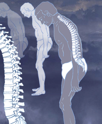
An illustration of ankylosing spondylitis, which commonly affects the joints between the spinal bones and the sacroiliac joints, which link the spine to the pelvis. The joints become painful and stiff. In severe cases, the vertebrae may fuse together completely. The condition is treated with painkillers and regular exercise.
Dominique Duval/Science Source
It was a shock to open the exam room door and shake hands with Joshua N., age 18. He reached up and pulled back a shock of stringy, coal-black hair. “It’s okay,” he said. “I’m used to the staring.” By skootching his bottom to the edge of the exam chair and extending his arms onto the arm rests, he was able to accommodate the curvature in his back and level his head. I met his eyes. We shook hands. I recalibrated and quietly asked him how long ago his back pain had begun.
“Maybe four, maybe five years ago.” His voice was silky and low, like a jazz radio host. It seemed disconnected from the frail young man in front of me. “A pillow?” he asked. He shifted slightly and grimaced. “Do you have a pillow you can put under my butt? Maybe another for my back? Chairs and I don’t get along so well.”
I reached into a lower cabinet and grabbed two pillows. Joshua N. half-stood, and I slid one pillow beneath him and placed the other low against the back rest. With half-closed eyes, he exhaled slowly and settled into the chair.
“So anyway, I saw my family doctor first. I guess we were living in Limington [Maine] then, and he didn’t know what to do with me. Eventually, he sent me on to orthopedics. The films didn’t show much back then. I mean, I didn’t have this f***ed-up, crippled look I do now. Orthopedics gave me some exercises and some pills. They basically said that I had a bad back, and I would have to learn to live with it. I got to the point that I stopped going to school. We lived two miles out on a dirt road, and the bus ride just about killed me. So I helped out around the house with my younger brother and sister, picked up kindling outside for the fire, learned how to cook—you know, tried to keep busy.
“I’ve been living on ibuprofen. It doesn’t help much, but when I run out, especially in the morning, I can barely move. They tried indomethacin a few years ago; I remember that by how much it messed up my stomach, but I guess that was better than the ibuprofen.”
“Have you tried anything else?” I asked.
“Last year, we moved up north to be closer to my mom’s family. She tried to home school me and took me to a lady who gave me a lot of herbs and vitamins. What a waste. Oh, here, the medical clinic gave me these two weeks ago.”
He handed me a vial of hydrocodone. “How often are you taking these?” I asked.
“A couple at night. I rest better, but feel wacky if I take them during the day. Can you refill it? I’m running low. Anyway, I’m in Portland [Maine] now, living with an older brother, and the medical clinic at Maine Medical Center says the X-rays show my back is messed up big time.” He smiled out of the corner of his mouth. “Big surprise there.”
I reached over and picked up the folder off the exam table and slid out the films. A surge of frustration and anger flowed over me: How had this poor guy slipped through the cracks? I put on my stethoscope and listened to Joshua’s lungs to settle my mind. It’s not often I’m shaken to the point that emotions take me out of my usual routine. I consciously made an effort to begin over. Where else has this disease settled in?
The Exam
“Besides the back, are you hurting anywhere else?”
“My knees are killing me. That’s been maybe a year or two. Can you help me roll my pants legs up?”
I pulled up the right jean cuff. A barbed-wire tattoo encircled one calf. Over the mid-shin was a crusty, oozing scab. It was a challenge to roll the jeans up and over the knees, so I had Joshua N. undo his belt and slip the jeans down. The sight was gut-wrenching. Each knee held enough fluid to fill a grapefruit. The fluid seemed to extend into the back of the calves—giving them a pseudo-Popeye appearance—and tracked upward into the lower thighs. Above and below the swelling, the muscles in the legs were atrophied. I placed one hand on the kneecap and the other on the back of the calf to assess range of motion.
“I’d appreciate it if you more or less, just look. They don’t move so well.” He draped the cloth gown I’d given him to cover himself. “It’s OK if you call me Josh. That’s my name.”
“Okay, Josh. Mind if I take off your sneakers?” With the enormous knee effusions, I was looking for clues of inflammation elsewhere.
Although nearly all rheumatologic conditions are more common in women than men, ankylosing spondylitis, at least in its most severe forms, occurs more frequently in men.
“That’s fine. I can’t reach down there anyway, so my brother scrubs them if he has time.” As I undid the Velcro fasteners and slid his shoes and socks off, I could see that two of the toes on the right foot were obviously red and swollen. The left ankle moved poorly and was tender to touch. When Josh tried to flex and extend each knee, the muscles in his thigh quivered.
“I haven’t been able to straighten either knee for, I don’t know how long.”
Now I understood why Josh was even shorter than what one would expect from the curvature of his back alone. With the knees partially flexed to accommodate the huge effusions, his hips would flex to compensate. Between the curvature of the back and the flexed knees and hips, Josh was 8–10 inches shorter than if he were fully erect. In truth, Josh resembled a modern-day Hunchback.
Above the waist, fluid within the elbow joints prevented full extension. One wrist was warm and boggy. His neck lacked any meaningful rotation or extension. When I asked Josh a question, he shifted his entire body to make eye contact; the neck was fused en bloc.
Rheumatologists have known for decades that patients who are HLA-B27+, may develop reactive arthritis after GI infections … [but] clarifying the triggers for AS has been elusive, in part due to the complex immunology of the gut.
Diagnosis
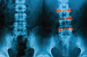
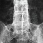
This L-S spine X-ray image, AP view, depicts a comparison of a normal (left) and ankylosing spondylitis (right) lumbar.
Suttha Burawonk/shutterstock.com
The diagnosis was evident on physical exam alone: ankylosing spondylitis. And I knew that even if medications could put out the fire of inflammation, reduce the swelling, improve the pain, there was no resetting the damage.
I stuck my head out the door and asked Joanne to set up for an aspiration and injection of each knee. On second thought, I decided to go through the details with her outside the room—and take a break. Josh was too crippled to lie flat on the exam table, and I’d need to position his knees carefully where he sat to drain them as completely as possible. I reminded her to bring in the entire aspiration tray: “We’ll need multiple 50 cc syringes, red-top tubes, and 2 cc of Depo-40 for each knee.”
Reflecting on Another Case
I had a few minutes. The next patient on my schedule had cancelled. I sat down in my office chair for a moment and drifted back to what I knew about ankylosing spondylitis (AS), and as usual, there were more unanswered questions than answers. For one, why is it that ankylosing spondylitis can be subtle in one patient and cruelly fulminant in another? Why are the sacroiliac joints involved (in most patients) early in the disease before inflammation ascends up the axial spine? Why do many AS patients have inflammatory eye disease with unilateral iritis? On colonoscopy, why do many AS patients have either macroscopic or microscopic patches of inflammatory bowel disease?
My thoughts drifted back to another young man with a similar duration of back pain, but an entirely different trajectory.
I met Michael T. in my first year of practice. Wearing wire-rimmed glasses and dressed in khakis and a button-down white shirt, he was of normal height and weight, and lacked the hang-dog slouching shoulders of chronic pain. He appeared fit, relaxed and comfortable in his own skin. The referral was from a neurosurgeon who had reviewed an MRI demonstrating low-grade congenital spinal stenosis. The stenosis was so mild it couldn’t possibly account for his painful, stiff back. Surgery was out, the surgeon told Michael T. Maybe I could shed some light.
“So you’ve had back pain for six years,” I noted as I reviewed his chart and looked over his normal plain films of the lumbar spine and pelvis. “Any injuries?”
“Oh sure, I mean I’ve been dinged up from time to time. Nothing serious. I fell off our roof shoveling snow when I was 16, then, fell again rock climbing a year later, but that was no big deal. I’ve never connected any injuries to my back pain; it’s just there. Every morning I wake up like Groundhog Day, and there it is, again.”
“Car accident? Litigation? Are you applying for disability?”
“Are you crazy?” he bristled. “I’m a physics major. My hero, Steven Hawking, has Lou Gehrig’s disease and can’t move his arms and legs, but the guy is rolling out mathematical solutions to stuff Einstein passed on. Listen, I’ve got a bad back. It’s not the end of the world, but I’ve tried all the stuff you guys tell me to do. Took the drugs, gone to therapy, stretched like crazy. Hey, my sister dragged me to her Yoga class, and that was cool. I felt good for maybe an hour. I don’t even know why they sent me here. Don’t rheumatologists see a bunch of old people with arthritis?”
“Okay. Sorry,” I said, ignoring the dig. “Let’s start over. Where exactly do you hurt?” Mike stood up and slid his hands around the lower lumbar region and sacrum. “Does this hurt?” I firmly palpated the para-lumbar muscles and the sacroiliac region.
“No,” he shrugged. “That’s the thing. When I reach back there, I can’t find a spot that’s particularly tender. It’s a deeper ache. Mornings are tough. I feel like the tin man.”
“Stiff?”
“Yeah, like major stiff. I shower and stretch before class, but if I sit for more than 10 minutes it all rolls back. So I’m constantly stretching, trying to loosen up, which is the only thing besides naproxen sodium that helps at all.”
“Can you touch your toes without bending your knees?” I squatted down behind him and placed my thumbs over the upper aspect of his sacroiliac joints. “Wait, I’m going to put two marks on your back before you bend forward.”
Michael reached for the floor, but his fingertips extended only to his mid-calves. “Not so good,” I noted. “Can you try that again? Keep your knees straight and give me your best effort.” This time, I measured the distance that his lumbar spine distracted between the two points I had marked. It barely budged. The test, a Schober’s maneuver, measured 1 cm. Clearly abnormal.
I hung the MRI up on the view box and agreed that the changes of congenital spinal stenosis were minimal. But where the MRI image extended into the sacroiliac joints was the answer. Both sides of the SI joints demonstrated definite changes of inflammation.
Both young men, Joshua and Michael, had ankylosing spondylitis. In one, the disease tore through the tissues like an unrelenting malignancy. In the other, the disease smoldered in place with only the subtlest of signs: MRI evidence of sacroiliac inflammation. Well, not quite. I had noticed that the left pupil was dilated slightly more than right, so I asked Michael if he had problems with the eye.
“You mean the iritis, which keeps coming back?” Sure. “My dad has the same thing. In fact, it’s the same eye. Weird, huh? But he’s lucky, he hasn’t ever had a shred of back pain. Lucky guy.”
Back to the Present Case
I re-entered the exam room and focused on the present task: Josh’s huge knee effusions. When I entered the right knee with a 16 gauge needle, there was so much pressure, my 50 cc syringe filled almost immediately. Joanne handed me another syringe, then another and another. Before I was done, six full syringes—300 cc—of amber fluid had been removed from the diseased knee. Another 250 cc was aspirated from the left knee, another record.
Even after the fluid had been aspirated from the knees, an audible grating, clunking sound occurred as I tried to flex and extend them. I hung up the films that Joshua had brought from Maine Medical Center. More bad news. From the pelvis to the neck, the spine was fused in a forward flexed position: a bamboo spine. The sacroiliac joints were fused. The hip joints were narrowed. For the first time in my practice career, I decided to admit a patient, an 18-year-old patient, for inflammatory arthritis.
In recent years, several studies have assessed whether altering the bacterial biome of the gut may lead to clinical improvement in ankylosing spondylitis. … But probiotics alone may not be the answer.
Ankylosing Spondylitis
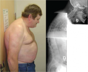
This 48-year-old man with ankylosing spondylitis has marked restriction in cervical spine range of motion. The composite of the cervical, thoracic and lumbar spine radiographs show syndesmophytes bridging adjacent vertebrae throughout the spine.
ACR IMAGE BANK
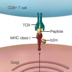
AS, unlike rheumatoid arthritis, undeniably goes back thousands of years and afflicts crocodiles, as well as humans.1 Although nearly all rheumatologic conditions are more common in women than men, AS, at least in its most severe forms, occurs more frequently in men.2 We know that the gene coding for the Class 1 MHC molecule, HLA‑B27, is found in more than 90% of Caucasians with AS, and that bacteria in the gastrointestinal tract in some poorly defined way, play a role in the pathogenesis of this disorder.
Rheumatologists have known for decades that patients who are HLA‑B27+ may develop reactive arthritis after GI infections due to Campylobacter, Salmonella, Shigella or Yersinia.3 After exposure, there is a delay of one or two weeks before an explosive large joint polyarthritis occurs (sometimes with axial spine and sacroiliac involvement). The joints themselves are not infected, but are the target of an immunologic storm of pro-inflammatory cytokines. In time, however, reactive arthritis usually resolves.
But what of AS? How important are bacteria in the pathogenesis of this more insidious and chronic disease? The clues are coming from both basic science and clinical research, but as yet there is not a comprehensive explanation for how we get from the Class I HLA-B27 cell surface molecule to inflamed joints, from a normal spine to a bamboo spine. How do we explain Joshua N or Michael T?
Clarifying the triggers for AS has been elusive, in part due to the complex immunology of the gut. We know that billions of bacteria coexist with us in the gut, but until recently, exactly what they were doing there was a complete mystery. Basic science is beginning to unravel the complex ecology of the gut, which should lead to a clearer understanding of how our own native bacteria affect health.
Scientists can now transfect an attenuated virus (a virus incapable of causing disease), and insert the human HLA-B27 gene into the virus’s genetic code. When the virus is injected into a mouse, copies of the human HLA-B27 markers can subsequently be detected on the cell surface of the mouse’s cells. What happens next to these previously normal mice? They develop diarrhea, inflammation in the bowel and skin, and after a delay of a week or so, inflammation in the joints. This study raises the question: Are the mice’s own native bacteria interacting with the human HLA-B27 gene and triggering arthritis in distant joints?4
To answer this, a second experiment was performed.5 Researchers gave the normal mice a combination of antibiotics to sterilize the bowel before they were transfected with the human HLA‑B27 antigen. And then the researchers waited. And nothing happened. They assessed the bowel, and there was no inflammation. They checked the skin and joints. Normal. It seems that in order to trigger widespread inflammation in HLA‑B27-positive mice, bacteria in the bowel are necessary to interact with the immune system.
But which bacteria? As long ago as 2002, I read with interest an article titled, “Klebsiella is the cause of ankylosing spondylitis.”6 After reading it, I was convinced that the authors had pinpointed the trigger for ankylosing spondylitis. To my chagrin, I flipped the page to the next article, titled, “Klebsiella is not the cause of ankylosing spondylitis.”7 In equally convincing terms, it refuted every argument that only minutes before had seemed irrefutable. So goes the search for truth.
In recent years, several studies have assessed whether altering the bacterial biome of the gut might lead to clinical improvement in AS. An open-label study that assessed the effect of treating AS patients with moxifloxin was published in 2007.8 This 12-week study of 76 patients demonstrated mild, statistically significant improvement in AS symptoms. But probiotics alone may not be the answer. A probiotic did not significantly benefit patients with ankylosing spondylitis compared with placebo in a controlled trial published in the Journal of Rheumatology in 2010.9
Hospital Visit
After hours, I stopped by to see Josh at Maine Medical Center. He was asleep after a chair shower, curled up on his side, his hair neatly combed, the T-shirt replaced by a hospital gown. An IV infusion of methylprednisolone sodium succinate was running in. Physical and occupational therapists had written their notes. Nursing had already called me regarding pain management. For the time being, Josh was receiving hydrocodone and indomethacin. I reviewed with his primary nurse the concept that Josh’s pain was different than, say, postoperative pain. Control of his inflammation should result in better control of his pain. We shouldn’t rely on narcotics.
“But he’s rating his pain as an 8 on a 1–10 scale,” she reminded me.
“I know, but let’s go easy on the narcotics.” She was not totally convinced.
I was already considering the next step, an infusion of infliximab, a TNF-alpha blocker. On his right forearm, nursing had already placed a PPD to ensure Josh had never been exposed to tuberculosis.
AS, unlike rheumatoid arthritis, undeniably goes back thousands of years & afflicts crocodiles, as well as humans.
Josh stirred. “I hated the idea of coming in the hospital, but this is okay. People here have been very nice.”
I sat down at the bedside and tried to explain ankylosing spondylitis in terms of what Josh could connect with. His last year of formal schooling was 9th grade, but he had a good grasp of the basics and was open to “anything that can help.”
The hospitalization was of some benefit. On the plus side, Josh was soon able to ambulate without a cane, and the swelling in his knees, ankle, elbow and wrists improved dramatically. The first infusion of infliximab went well. His pain was better controlled; he no longer felt sedated or loopy with daytime use of hydrocodone.
But there were plenty of negatives. When he vomited up blood on Day 3, a stomach ulcer was demonstrated on endoscopy, and we needed to stop his anti-inflammatory, indomethacin. X-rays of his knees demonstrated that the cartilage was nearly gone. At age 18, he was bone on bone and looking at early bilateral knee replacement. The hips were nearly as bad. His neck and back regained zero motion. He remained stooped, lacking horizon vision, perpetually gazing at his shoe tops.
In the Long Term
I knew that we were dealing with a particularly aggressive form of AS when I saw Josh only two weeks after discharge and the fluid in his knees had recurred. Markers of inflammation—ESR and CRP—were nearly back to their prehospitalization levels. He was using a cane again. I titrated up the amount of infliximab for the next infusion and once again tapped and injected the knees.
In the ensuing weeks and months, Josh’s disease slowly came under some semblance of control. I was able to taper down the prednisone and cautiously restarted the anti-inflammatory, celecoxib, combined with omeprazole to prevent recurrence of his gastric ulcer. But Josh’s need for the hydrocodone accelerated. From time to time, he called for early refills, and on one occasion, he claimed that his pills were ruined when the top of his prescription vial came off and the pills were soaked in the rain. He missed an appointment and then another. But he always showed for his infusion of infliximab. It worked, and he knew it.
A year after our first visit, I admitted him with pneumonia, which was slow to resolve. On my way into the hospital one early morning on Day 4 of his hospitalization, I watched Josh wheel an IV pole out to the smoking area outside the hospital and light up. Confronting him, he promised he would try to quit. Out of safety concerns, I held his infliximab for a month, and the AS promptly flared. More aspirations and injections followed. Back on track with the infliximab, his disease improved, somewhat.
At some point, Josh stopped coming to the office. He didn’t refill his prescriptions. We called his cell phone; no answer. I wondered if he’d moved or committed suicide, or overdosed. We never had been able to titrate the hydrocodone down. His burden of pain was real; between his end-stage knees and painful back, it seemed appropriate for Josh to be treated with narcotics. But I had my concerns. I checked the obituaries in the paper. Joanne finally tracked down Josh’s mother who was back living in northern Maine. I called her.
“Josh is in jail,” she said. “Dealing drugs. They got him, and now he’s in Thomaston.” There was a long pause. “I can only hope they give him a break. That boy has suffered enough.”
I made a few calls. Through the prison system, Josh was getting a reasonable alternative to the intravenous infliximab, etanercept, which could be administered as an injection. I spoke to the doctor in charge, who reassured me that Josh was getting good care.
As the months and then years flowed by, I often thought of Josh and wondered if he’d ever had his knees replaced. I never did aspirate more fluid from a diseased knee. Josh’s aspirate of 300 cc remained a dubious record. At our weekly rheumatology conferences, Josh’s name would come up, as in, “This patient has very active disease, but not as severe as Josh N.’s ankylosing spondylitis.” The other rheumatologists would nod their heads and agree. No, Josh was the worst. The absolute worst. Whatever happened to him?
Then one day, Josh came in for an office visit. By now, he was in his mid-20s and had suffered with AS for 12 years. I watched as Joanne guided him down the hallway to the scale. Inside the exam room, he told me that his five years in prison was not altogether wasted. He’d gotten his GRE and learned a trade. A local vegetarian restaurant had recently hired him as a line cook. I watched Josh arrange himself in the exam chair—one pillow behind his back and the other under his butt. The T-shirt had been replaced by a loose-fitting, floral bowling shirt. My eyes scanned down to the front pocket: No cigarettes. Good. On his feet he wore bowling shoes with a Velcro strap closure. Very classy.
Pulling up a pant leg, he showed me the vertical scar of matching knee replacements. Beneath his shirt, he wore a soft Velcro back brace, which helped, he said, but most of the time he just put the pain aside and tried to stay busy.
“It’s great to see you, Josh,” I began. “I see that you’re still on the etanercept they switched you to in Thomaston. Is that working well enough?”
“Well enough,” he said.
I scratched my cheek, waiting. If he was after narcotics, I was ready to say no, but Josh had other ideas. “I’ve been reading about a big surgery. They break your back in multiple places, put in some rods and screws—well, maybe more than a few rods and screws—and straighten the back. What do you think?”
I knew what Josh was referring to. It was a monster of a surgery. Only specialized centers had spine surgeons with experience enough to break up a bamboo spine and put it back together again. This was much more than corrective surgery for scoliosis. Josh would have to go out of state for the consult. Even then, there was no guarantee that a surgeon would take him on. The risk of infection, bleeding, hardware loosening or even paralysis was very high. If he survived the surgery, he could go through months of rehab and be worse off than he was now.
“You sure you want to start down this road?” I asked.
“Positive.” Josh stood up and found his cane. His eyes locked onto his shoes. “I know where I’ve been. I just want to see where I’m going. That’s not too much to ask, is it?”
I began the paperwork.
Charles Radis, DO, is clinical professor of medicine at the University of New England, College of Osteopathic Medicine and employed part time at Maine Coast Memorial Hospital in Ellsworth, Maine.
References
- Braun J, Sieper J. Ankylosing spondylitis. Lancet. 2007 Apr 21;369(9570):1379–1390.
- Moodie R. Palaeopathology. An Introduction to the Study of Ancient Evidences of Disease. Chicago and Urbana: University of Illinois Press; 1973:128.
- Yu D, Kuipers JG. Role of bacteria and HLA-B27 in the pathogenesis of reactive arthritis. Rheum Dis Clin North Am. 2003 Feb;29(1):21–36, v-vi.
- Hammer RE, Maika SD, Richardson JA, et al. Spontaneous inflammatory disease in transgenic rats expressing HLA-B27 and human beta-2m: An animal model of HLA-B27 associated human disorders. Cell. 1990 Nov;63(5):1099–1112.
- Tautog JD, Richardson JA, Croft JT, et al. The germfree state prevents development of gut and joint inflammatory disease in HLA-B27 transgenic rats. J Exp Med. 1994 Dec 1;180(6):2359–2364.
- Ebringer A. Ankylosing spondylitis is caused by Klebsiella. Evidence from immunogenetic, microbiologic, and serologic studies. Rheum Dis Clin North Am. 1992 Feb;18(1):105–121.
- Russell AS, Suarez A. Ankylosing spondylitis is not caused by Klebsiella. Rheum Dis Clin North Am. 1992 Feb;18(1):95–104.
- Ogrendik M. Treatment of ankylosing spondylitis with moxifloxacin. South Med J. 2007 Apr;100(4):366–370.
- Jenks K, Stebbings S, Burton J, et al. Probiotic therapy for the treatment of spondyloarthritis: A randomized controlled trial. J Rheumatol. 2010 Oct;37(10):2118–2125.
