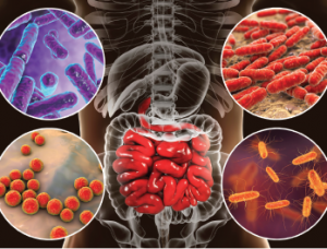
Bacteria (e.g., Bifidobacterium, Lactobacillus, Enterococcus and Escherichia coli) colonize different parts of digestive system.
Kateryna Kon / shutterstock.com
Evidence is accumulating that the microbiome may be an important part of the pathogenesis of many autoimmune diseases. Two recently published articles report on how translocation of the gut bacterium Enterococcus gallinarum drives autoimmunity in mice and humans, and on the role of other commensal bacteria in triggering immune responses—specifically to the autoantigen Ro60, which is targeted in systemic lupus erythematosus (SLE).1,2
“Both of these studies deal with the role of the microbes that live in our gut and other places of the human body of patients with autoimmune disease,” says Martin A. Kriegel, MD, PhD, adjunct professor of immunobiology at Yale University School of Medicine, New Haven, Conn., and lead author of the two reports. “The overarching theme in my laboratory is the role of the microbiome in autoimmune diseases. These results work toward a better understanding of how the microbiome fits into the development and treatment of these diseases.”
Indication of Bacterium Translocation
The study published in the March edition of Science looked at indicators of translocation of E. gallinarum in a hybrid autoimmune mouse model under pathogen-free conditions.1
The researchers studied the response in these mice to classic autoantigens in SLE as well as endogenous retrovirus glycoprotein 70, which had previously been shown to drive lupus kidney disease in this model. Mortality, lupus-related autoantibodies and autoimmune manifestations were all lowered after oral administration of antibiotics. In addition, translocation of the microbiota was successfully suppressed by vancomycin or ampicillin. Both also prevented mortality by autoimmune disease in this mouse model.

Dr. Kriegel
Uptake of an orally fed fluorescent dye into systemic circulation of the pathogen-free mice indicated impaired gut barrier function when compared with another mouse line that was not genetically predisposed to autoimmune disease. The researchers were able to detect bacterial growth in the mesenteric veins, mesenteric lymph nodes (MLN), spleen and liver, further indications of translocation.
When the group looked at full-length ribosomal DNA sequencing of single colonies from aerobic and anaerobic MLN, liver and spleen cultures, E. gallinarum predominated in all tissues including in the mesenteric veins of 82% of the lupus-prone mice. It was also visualized in situ by fluorescence within MLN and the liver. Control mice showed no bacterial growth.
Species-specific polymerase chain reaction (PCR) did not detect E. gallinarum DNA in stool samples of human or murine autoimmune hosts. However fecal or mucosal cultures followed by species-specific PCR consistently showed the bacterium in the feces, small intestine and liver of the hybrid mice, suggesting that some disease-relevant gut microbes are not easily detectable in conventional stool studies because they hide within the body.
“This demonstrates that there is leakiness not only in inflammatory bowel disease, where we know about gut barrier dysfunction, but also in SLE and autoimmune hepatitis, where there is no overt inflammation in the gut,” says Dr. Kriegel. “We saw this based on markers for leakiness in the stool of autoimmune patients. Importantly, we also found E. gallinarum-specific DNA in liver biopsies taken from these patients.”
‘The overarching theme in my laboratory is the role of the microbiome in autoimmune diseases. These results work toward a better understanding of how the microbiome fits into the development & treatment of these diseases.’ —Dr. Kriegel
Can Bacteria Produce Pro-Inflammatory Pathways?
The researchers next addressed the issue of whether E. gallinarum could induce pro-inflammatory pathways in small intestine tissue during translocation. They also wanted to know if the bacterium could alter the gut barrier.
To do this, mice monocolonized with E. gallinarum were compared with mice monocolonized with two other gut bacteria. RNA expression profiling was used as one of the readouts.
The presence of E. gallinarum was shown to downregulate ileal molecules related to barrier function in the gut, the mucosal layer and antimicrobial defense. It was also noted that monocolonization with E. gallinarum upregulated those related to inflammation and induced systemic autoantibodies. One of the upregulated markers was Enpp3, a molecule known to increase key cells contributing to the type 1 interferon (IFN) signature in human SLE.
Another link to disease was found when a vaccine for E. gallinarum was introduced. Translocation in autoimmune-prone mice induced autoantibodies and caused mortality. Both were prevented by vaccination.
“An important part of the study is not only that we were able to associate E. gallinarum with autoimmune diseases,” says Dr. Kriegel. “We also did mechanistic work in the test tubes and animals that linked these bacteria to the pathways that are detrimental to patients with autoimmune disease.”
Gut Leakage & Human Disease
Turning to an analysis of gut leakage and effects on human disease, longitudinal analysis of stool from SLE patients revealed impaired gut barrier function with an increase in fecal albumin and calprotectin. The researchers then tested for E. gallinarum translocations in human livers of patients with SLE and autoimmune hepatitis (AIH).
Liver biopsies from three lupus patients were positive for E. gallinarum. Of the six control biopsies from healthy liver transplant donors, four were positive for other Enterococcus species, but not E. gallinarum.
Sterile liver tissue samples from the same control donors were compared with tissues from AIH and cirrhosis patients. Cirrhosis patients were selected as a positive control because they are known to have grossly impaired gut barriers.
Studies of rDNA sequencing and PCR showed E. gallinarum predominated in diseased tissue. The majority of AIH biopsies, but not the healthy controls, was positive for this bacterium. There were also enhanced adaptive immunity responses to E. gallinarum because the majority of AIH and SLE patients showed increased antibody titers against it and, particularly, its RNA. The latter could act as a cross-reactive stimulus to drive immune deposits in lupus kidney disease.
“This shows that the bacterium triggers immune reactions and pathways of the immune system that are important in autoimmunity such as those leading to Th17 cells,” says Dr. Kriegel. “We have found that certain bacteria that cross the gut barrier induce cytokines and immune pathways leading to inflammation.”
Ro60 Autoantigen as a Trigger for SLE
The other article appeared in Science Translational Medicine. Dr. Kriegel, together with co-lead author Sandra L. Wolin, MD, PhD, from the RNA Biology Laboratory at the National Cancer Institute and professor emeritus at Yale, and associates looked at orthologs of the Ro60 autoantigen as possible triggers for autoimmune responses in SLE.2
“The earliest antibodies in lupus are directed against the RNA binding autoantigen Ro60, but the persistent triggers against the evolutionarily conserved antigen remain elusive,” Dr. Kriegel wrote in the article. “We identified Ro60 orthologs in a subset of human skin, oral and gut commensal bacterial species and confirmed their presence in patients with lupus and healthy controls. We hypothesized that Ro60 orthologs may trigger autoimmunity via cross-reactivity in genetically susceptible individuals.”
Sera from human anti-Ro60-positive SLE patients were found to bind to bacterial Ro60 proteins. CD4 memory T cell clones for the human Ro60 autoantigen were activated by proteins and peptides from skin and mucosal Ro60-containing bacteria. This, according to the authors, is supportive of T cell cross-reactivity in humans.
Spontaneous Anti-Human Ro60 Antibody Production
Further indications were seen when germ-free mice monocolonized with one Ro60-containing bacterium spontaneously began producing anti-human Ro60 T and B cell immune responses. The researchers also developed glomerular immune complex deposits after monocolonization. This suggested a linkage between Ro60 responses in vivo, with production of Ro60 autoantibodies coinciding with signs of autoimmunity.
“Both of these studies deal with the role of the microbes living in our gut and other places, like the skin, in autoimmune diseases, such as SLE,” says Dr. Kriegel. “One important takeaway from the translocation study is that there are certain circumstances in which bacteria can get into our living tissues without being an infectious agent in the usual way. They don’t make us sick as pathogens do, but live within us and trigger autoimmune pathways.”
Biomarkers & Treatments Possible
The researchers also think these findings may make it possible to develop biomarkers. Then it might be possible to develop treatments that stop SLE and other autoimmune diseases early enough to avoid major damage.
“We have done interventional studies in animals, and there were two important findings in this context,” says Dr. Kriegel. “One is that certain oral antibiotics were able to prevent the disease in the animal model. Second, we developed a vaccine against E. gallinarum that depleted the bacteria from the tissues and prevented the occurrence of autoimmune disease in mice.”
Kurt Ullman has been a freelance writer for more than 30 years and a contributing writer to The Rheumatologist for 10 years.
References
- Manfredo Vieira S, Hiltensperger M, Kumar V, et al. Translocation of a gut pathobiont drives autoimmunity in mice and humans. Science. 2018 Mar 9;359(6380):1156–1161.
- Greiling TM, Dehner C, Chen X, et al. Commensal orthologs of the human autoantigen Ro60 as triggers of autoimmunity in lupus. Sci Transl Med. 2018 Mar 28;10(434). pii: eaan2306.


