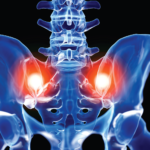
 Radiologists and rheumatologists who interpret pediatric pelvic magnetic resonance imaging (MRI) scans must be able to differentiate inflammatory changes from the normal physiologic changes of a maturing sacroiliac (SI) joint. However, this ability varies by specialist, a point highlighted by a new study from Pamela F. Weiss, MD, associate professor of pediatrics and epidemiology, University of Pennsylvania Perelman School of Medicine, Philadelphia, and colleagues. Their findings suggest additional training may be needed for radiologists regarding the appearance of the maturing SI joint on MRI.
Dr. Weiss and colleagues evaluated the agreement between local radiologists’ interpretations of pediatric SI joint MRIs and a central imaging team’s interpretation. Their study was published in the June issue of Arthritis Care & Research.1
Study Details
The study included the evaluation of the SI joints of 120 children performed in eight pediatric hospitals across North America. With a median age of 14 years, the children had a median disease duration of 0.8 years at the time of imaging. Half the children were male, and the majority were white (76.8%). In addition, the majority of children had a diagnosis of enthesitis-related arthritis (82.4%). The investigators coded the global impression of sacroiliitis based on the impression section of the radiology report. If the impression did not differentiate between active inflammation and chronic sacroiliitis, the researchers looked at the findings section of the radiology report. If bone marrow edema in or along the SI joints was noted in the findings section, then the report was coded as chronic inflammation.
The MRIs were independently reviewed by three experienced musculoskeletal pediatric radiologists serving as the central review team for the study. These three radiologists were selected for their ability to identify inflammatory and structural lesions at the SI joint. The majority of the time (84%), the central radiologists agreed on the presence or absence of active inflammation and chronic lesions. The results of the central review team were compared with reports from the children’s local radiologists. The authors note that no standardized assessment for local interpretation exists, and the imaging quality of the studies reviewed was highly variable—even within the same institution.
The local reports had a low, positive predictive value of 51.8%. In other words, only 51.8% of the diagnoses of sacroiliitis made locally were confirmed by the centralized readers. Only rarely (2% of the time) did the central radiologists identify active inflammation when the local radiologists did not identify active inflammation. It was much more common (22.5% of the time) for the local radiologists to identify active inflammation and the central radiologists conclude that there was no active inflammation. Thirteen of the studies were missing coronal oblique sequences, making visualization of the synovial section of the joint more difficult than if the sequences were available. However, when investigators performed a sensitivity analysis that excluded all studies with any quality issues or missing coronal oblique sequences, the rates of discordance were approximately the same.
When the researchers performed a second sensitivity analysis using only cases with total central radiologist agreement (84% of total cases), the overall diagnostic test statistics were unchanged for active inflammation with a positive predictive value of 52.4% and a negative predictive value of 95.9%, meaning there was a 52.4% probability that patients identified as positive for active inflammation by local reports were positive and a 95.9% probability that patients identified as negative for active inflammation by local report were negative. When researchers performed this second sensitivity analysis for the presence of chronic lesions, the positive predictive value was slightly improved, to 70.0%, whereas the negative predictive value was similar at 81.9%.
The authors concluded from this second sensitivity analysis that most of the discordance appeared to be due to errors in differentiating normal physiologic metaphyseal equivalent signal from pathologic subchondral marrow edema. The investigators found that in 27 cases (23%), local radiologists identified active inflammation when the central review reading deemed the same patients’ joints as normal.
Implications for Patients
Dr. Weiss calls this an important study, noting that, “because this is a real problem for the evaluation of these patients, we want interpretations to be as accurate as possible.”
She also emphasizes this study represents a concerted effort across eight institutions in the U.S. The study revealed that not all centers use the same sequences, and many do not perform the coronal oblique sequences, which are ideal for visualizing the synovial part of the joint. Although this situation is a problem, it’s not the major problem.
“This is not an image quality issue,” says Dr. Weiss. “This is an interpretation issue. … There are some normal maturational changes that occur at the SI joint that can be confused for minor inflammation.”
Dr. Weiss says the conclusions of the paper are clear: It’s challenging to interpret the MRI of the SI joint in children if you aren’t familiar with the appearance of a normal maturing pelvis.
On the positive side, local radiologists are quick to recognize and diagnose SI joint inflammation; very few incidents were missed by the local radiologists. Instead, the study points to a problem of misidentifying normal maturing SI joints as sacroiliitis. When radiology reports indicate findings consistent with active sacroiliitis, the rheumatologist may be influenced toward treatment with a biologic.
In 88% of the MRIs reviewed, the treating rheumatologist’s interpretation of the imaging, as documented in the medical records, agreed with the local radiology report, suggesting that they were transcribing the local radiologist’s report into their notes. Therefore, Dr. Weiss emphasizes the importance of accurately diagnosing patients to avoid both under- and overtreatment.
“We are right in the vast majority of cases,” says Dr. Weiss. “We just want to be right in all of the cases.”
SI joint inflammation is just one cause of low back pain. Often, physical therapy rather than biologic therapy is indicated. Because rheumatologists are making this important decision based on MRI reports, MRI interpretations must be as accurate as possible.
Solutions
According to Dr. Weiss, a big contributor to this problem has been the lack of materials to familiarize radiologists with the normal maturational changes that occur in the SI joint. However, in July 2021, radiologists published an atlas of pediatric structural changes. The atlas uses the preliminary, updated JAMRIS (Juvenile Idiopathic Arthritis MRI Score) scoring system and is intended to serve as a reference for the assessment of structural lesions in patients with SI joint arthritis.2
Dr. Weiss believes these resources will help familiarize a wider audience with the appearance of the normal pediatric SI joint because “images and examples are invaluable. … I am very optimistic things are going to improve with time and awareness,” she says.
For radiologists and rheumatologists, Dr. Weiss recommends an additional resource: the educational tools created by Care Arthritis, a company focused on the development of new imaging and biomarker technologies to advance personalized medicine for patients with arthritis. Although, according to Dr. Weiss, the Care Arthritis site includes primarily adult images, the free resource also has pediatric images.
Lara C. Pullen, PhD, is a medical writer based in the Chicago area.
References
- Weiss PF, Brandon TG, Bohnsack J, et al. Variability in interpretation of magnetic resonance imaging of the pediatric sacroiliac joint. Arthritis Care Res (Hoboken). 2021 Jun;73(6):841–848.
- N Herregods, WP Maksymowych, L Jans, et al. Atlas of MRI findings of sacroiliitis in pediatric sacroiliac joints to accompany the updated preliminary OMERACT pediatric JAMRIS (Juvenile Idiopathic Arthritis MRI Score) scoring system: Part II: Structural damage lesions. Semin Arthritis Rheum. 2021 Oct;51(5):1099–1107.

