The diagnosis of infective endocarditis is based on the modified Duke criteria.8 Although a definitive diagnosis can be provided only after histologic and microbial analysis of a vegetation or cardiac specimen, a clinical diagnosis of possible infective endocarditis can be made using the Duke criteria with the presence of one major and one minor criterion or of three minor criteria. The two major criteria are:
- A positive blood culture; and
- Evidence of endocardial involvement (an echocardiogram demonstrating a vegetation, an abscess or new valvular regurgitation).
The five minor criteria are:
- A predisposing condition;
- A fever;
- Vascular and embolic phenomena (including arterial emboli, septic pulmonary infarcts and Janeway lesions);
- Immunological phenomena (including glomerulonephritis, Osler nodes and rheumatoid factor); and
- Microbiological evidence of infection that does not satisfy the major criterion.
A vegetation is defined as an oscillating intracardiac mass on a valve or supporting structures, in the path of regurgitant jets or on implanted material in the absence of an alternative anatomic explanation.
Our patient satisfied one major criterion: endocardial involvement with an echocardiogram demonstrating a mitral vegetation. In addition, she satisfied three minor criteria: a predisposing condition of dental infection and dental surgery preceding her presentation, embolic phenomenon of splinter hemorrhages in the digits and immunological phenomena of glomerulonephritis with reduced complement levels and an elevated rheumatoid factor. Therefore, our patient would meet the criteria to assign a clinical diagnosis of possible infective endocarditis.
On the other hand, no well-established diagnostic criteria exist for the diagnosis of ANCA-associated vasculitis or GPA. Many physicians refer to the 1990 ACR classification scheme, which contains four criteria: nasal or oral inflammation, abnormal chest radiograph (with nodules, infiltrates or cavities), abnormal urinary sediment (with hematuria or red cell casts) and granulomatous inflammation on biopsy. The presence of two or more criteria yields a sensitivity and specificity of 88.2% and 92.0%, respectively for GPA.9 However, these criteria were developed prior to the routine use of ANCA and are not recommended for diagnostic purposes.
The diagnosis of GPA is typically made using clinical judgment by evaluating a patient’s symptoms together with laboratory and imaging findings while excluding other possible diagnoses, and by observation of improvement on immunosuppressive therapies.10
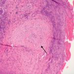
Our patient had many signs and symptoms that would suggest GPA, including a purpuric rash, hemoptysis and pulmonary hemorrhage resulting in respiratory failure and renal failure with hematuria. Her laboratory findings of an elevated ESR and CRP, and positive C-ANCA and PR3 antibodies, as well as lung opacities seen on chest X-ray and CT scan, were also all consistent with GPA. Her periungual skin biopsy contained thrombotic lesions more consistent with embolic phenomena from endocarditis rather than a vasculitis. However, the renal biopsy demonstrated a necrotizing glomerulonephritis with crescents and without immune complex deposits, which strongly favored GPA over infective endocarditis.

