Findings/Diagnosis
A posteroanterior supine radiograph of the sacroiliac (SI) joints (see Figure 1) demonstrates bilateral symmetric erosions of the SI joints without ankylosis and mild reactive sclerosis of both iliac bones adjacent to the joints (black circles). Review of thoracic and lumbar spine MRI examinations obtained two years prior to presentation show scattered areas of vertebral body bone marrow edema, centered at the vertebral body endplates and pedicles in the thoracic spine (see Figure 2, arrows) and in the anteroinferior corner of the L5 vertebral body in the lumbar spine (see Figure 3, arrow). Although initially interpreted as nonspecific, the MRI findings are consistent with spondylitis.
Based on the previous MRI findings of spondylitis and the current radiographic presence of symmetric erosive sacroiliitis, a diagnosis of ankylosing spondylitis was made. Subsequent HLA-B27 testing was positive, and the patient was started on therapy.
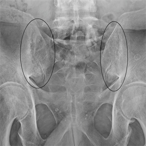
Figure 1
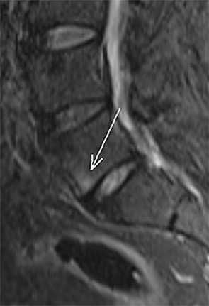
Figure 2
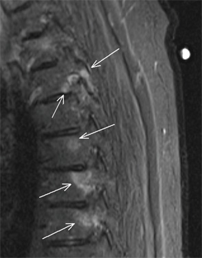
Figure 3
Jennifer L. Demertzis, MD, is a musculoskeletal radiologist at the Mallinckrodt Institute of Radiology at Washington University School of Medicine in St. Louis, Mo. She is excited to collaborate on this new feature in the journal and looks forward to seeing future cases contributed by readers.
