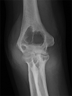
Figure 1: Right elbow anteroposterior radiograph

Figure 2: Right elbow lateral radiograph
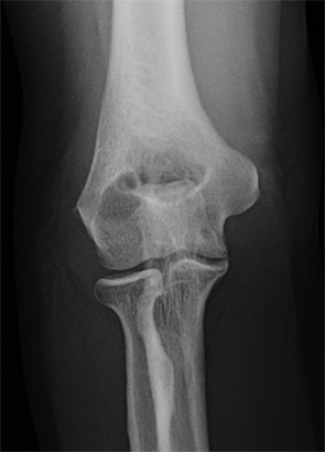
Figure 3: Right elbow anteroposterior radiograph, one year prior to current presentation
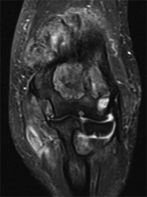
Figure 4: Right elbow coronal T2-weighted fat-suppressed MR image
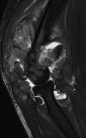
Figure 5: Right elbow sagittal T2-weighted fat-suppressed MR image
History
These images are of a 54-year-old woman with right elbow pain.
