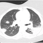
Many patients come to clinical attention based on imaging of the chest, which may demonstrate relatively symmetric and diffuse involvement of mediastinal and hilar lymph nodes, as well as thickened bronchovascular bundles. Unilateral pulmonary masses are rare, as are pleural effusions. When bronchoscopy is indicated, bronchoalveolar lavage (BAL) in patients with sarcoidosis would be expected to be predominantly lymphocytic.
Dr. Sharp pointed out that, in her clinical practice, she almost never orders a serum angiotensin-converting enzyme (ACE) level because the test is neither sensitive nor specific.
In the Organs
Sarcoidosis can affect nearly any organ in the body, and its presentations are often myriad. Cranial neuropathies can occur with involvement of the central nervous system, and Dr. Sharp has seen patients come to attention with the onset of acute hearing loss or dizziness.
Patients may also present with symptoms that mimic those of meningitis, encephalitis or transverse myelitis. If magnetic resonance imaging (MRI) of the brain is indicated to evaluate for evidence of leptomeningeal enhancement or other features of neurosarcoidosis, such imaging must be conducted with contrast to not miss these findings.
With regards to ocular involvement, uveitis is the most common finding. Cardiac involvement may include evidence of conduction delay, atrioventricular nodal block, bundle-branch block or fragmented QRS complexes.4 Cutaneous involvement of sarcoidosis can include classic findings, such as erythema nodosum or lupus pernio, as well as a wide range of skin lesions that have been described in association with the disease.
Patient Evaluation
Often, Dr. Sharp pursues all of the following tests when evaluating patients with sarcoidosis: computed tomography (CT) imaging of the chest, pulmonary function testing with spirometry and DLCO measurement at a minimum, electrocardiogram (ECG), complete blood count with differential, complete metabolic panel, measurement of calcium, 25-vitamin D, 1,25-vitamin D and referral to ophthalmology.
For assessing myocardial involvement, Dr. Sharp finds cardiac positron emission tomography (cardiac PET) more helpful than cardiac MRI because the latter cannot as easily distinguish between active disease and scarring. Even when directed at the heart, PET may pick up subclinical inflammation in other organs, such as the lungs or the liver.
Dr. Sharp explained that, although we typically think of patients with sarcoidosis as having restrictive or fibrotic lung disease, many other phenotypes are possible and should be screened for carefully. In a study of more than 600 patients with sarcoidosis, Dr. Sharp and her colleagues found that more than 50% had abnormal pulmonary function testing and that this finding included 22% with evidence of obstructive lung disease, 16% with an isolated reduction in DLCO and 15% with combined obstructive-restrictive lung disease.5