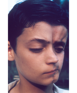
This was one of the very few slides in the first collection that included a distracting background. It was allowed in the collection only because the condition was so rare that no other slides were available (then called en coup de sabre lesion of linear scleroderma; today it would be described separately as Parry-Romberg syndrome, a distinction not made in the early 1970s). The photograph was submitted by Bernard Rogoff.
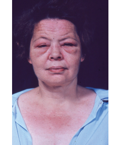
This photograph of a woman with dermatomyositis was included not only because it illustrates the rash and periorbital erythema, but also as an inside joke: John Decker, who closely advised the committee, rejected many slides because they showed periorbital edema, which he insisted was not necessary for a diagnosis of dermatomyositis. The committee challenged him to find a slide that did not show periorbital edema, and this is the best he could produce. Case closed.
Three years later, Barbara Ansell, MD, a pioneer in rheumatology and founder of pediatric rheumatology, wrote a review of the second edition that was published in the Journal of Clinical Pathology, calling it “well thought out and presented; in particular the accompanying photographs and descriptions are excellent.”
 Sidney Block, MD, a rheumatologist whose private practice encompasses northern and eastern Maine, chaired the Audiovisual Aids Subcommittee between 1982 and 1989. A tradition started during his tenure may be what many people today know best about the Image Library—the slide competition. Initiated in 1983, the annual contest was designed to expand the image collection by encouraging submissions from the entire rheumatology community. Prior to this, slides had been submitted primarily by committee members who either took the photographs themselves or sought them from other sources. There was always one member from the Mayo Clinic who was able to tap into that institution’s collection.