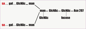
Many immunologists have theorized that autoimmune disorders result from aberrant function of the idiotypic network, and that IVIg contains antiidiotype antibodies that restore regulation by this pathway. Since immune responses to foreign antigens need to be self-limited, the idiotypic network has been postulated as a way to downregulate these immune responses specifically. According to this hypothesis, the variable regions of antigen receptors on T cells (TCR) and immunoglobulins contain unique amino acid sequences that determine their antigen specificity. This region is called the idiotope; all TCRs or antibodies that share an idiotope belong to the same idiotype. When a foreign antigen enters the system, one or a few clones of lymphocytes respond, and their idiotypes are expanded. Along with the expansion of particular idiotypes, antiidiotype responses are also triggered. These antiidiotypes can in turn downregulate responses by shutting off the antigen-specific or idiotype-expressing lymphocytes.1
According to this idea, antiidiotypic antibodies present in IVIg can bind to either surface immunoglobulin on B cells or circulating antibodies in plasma. IVIg can therefore ameliorate autoimmune disease by directly inhibiting autoantibody binding to its target antigen; it may also target autoreactive B-cell clones expressing or secreting the autoantibody for destruction. One disease that serves as a model for this proposed mechanism is acquired hemophilia caused by inhibitory autoantibodies to Factor VIII. Autoantibodies to Factor VIII may arise in nonhaemophilic patients with autoimmune disease, such as rheumatoid arthritis or systemic lupus erythematosus, as well as otherwise healthy individuals. Administration of high-dose IVIg in patients with acquired hemophilia can result in a rapid and prolonged depression of anti-VIIIc antibody titers. Further suggesting that antiidiotype antibodies are the active component of IVIg, subsequent studies showed that infusion of only the antigen-binding fragments of IVIg can reduce levels of the pathogenic antibody.8

An additional way that IVIg may modulate inflammation is the accelerated degradation of pathogenic IgG by saturation of the neonatal Fc receptor (FcRn), so named because it was initially identified in the neonatal intestinal epithelium. This IgG receptor aids in the transfer of IgG from mother to fetus and also protects IgG from degradation. FcRn is known to exist in many adult tissues, including intestinal epithelium, muscle, and skin. In states of hypergammaglobulinemia, (e.g., following high-dose IVIg), the FcRn is presumably saturated and the catabolism rate of the pathogenic antibody is accelerated. In murine models of autoimmune-mediated skin blistering diseases (e.g., bullous pemphigoid) administration of high-dose IVIg drastically reduces pathogenic IgG levels and prevents blistering. In contrast, mice deficient in FcRn are resistant to the development of blistering in this model and administration of high-dose IVIg does not confer any additional protective effect.9


