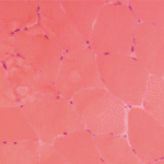
The inhibitory properties of IVIg may also result from its ability to scavenge active complement proteins, and studies have shown that high levels of IgG can inhibit the uptake of C3b and C4b onto the surface of sensitized guinea pig and human erythrocytes and prevent complement-mediated tissue damage. It is thought that IVIg binds the activated complement components C3b and C4b and therefore prevents their deposition onto target surfaces.10,11 A disease model in which IVIg convincingly modulates complement is dermatomyositis (DM). Dermatomyositis is an inflammatory myopathy mediated by the deposition of complement and formation of the C5-9 membrane attack complex (MAC) on intramuscular capillaries, leading to loss of capillaries and muscle ischemia and necrosis. Marinos Dalakas and colleagues from the National Institute of Neurological Disorders and Stroke evaluated whether high-dose IVIg can inhibit complement activity in a series of patients with DM treated with IVIg. They confirmed that IVIg reduces C3 uptake onto antibody sensitized erythrocytes.12 In addition, patients clinically improved and repeat muscle biopsies revealed disappearance of C3 and MAC deposits and improvement of the muscle architecture with an increase in muscle fiber size and number of muscle capillaries.12,13
In recent years, there has been a revolution in our understanding of the antiinflammatory effect of IVIg, with the important discovery that IVIg can be separated into extensively sialylated and nonsialylated IgG fractions. Essentially all of the effector functions of IgG, including the Fc-binding activity and the protective activity against pathogens, reside in the nonsialylated fraction. The sialylated fraction, comprising only 5% or less of the IgG pool, appears to contain all of the antiinflammatory activity. In murine models of inflammatory arthritis, administration of high-dose IVIg protects mice from developing joint inflammation. IVIg pretreated with neuraminidase to remove the terminal sialic acid residues, however, does not exhibit antiinflammatory activity. Conversely, in the experiments in mice, a sialic acid–enriched IVIg preparation showed a 10-fold enhancement in protection so that near equivalent protection was obtained at one-tenth the dose of unfractionated IVIg.14 An attractive possibility for developing new and more potent treatment would be a recombinant IVIg preparation that is composed of hypersialylated IgG molecules. (See Figure 2)
The antiinflammatory activity of the sialylated IgG is now thought to be mediated by binding of this type of IgG to SIGN-R1 (specific ICAM-3–grabbing nonintegrin-related 1) that is expressed on splenic macrophages in mice, or the human orthologue DC-SIGN (dendritic cell–specific intercellular adhesion molecule-3-grabbing non-integrin) expressed on dendritic cells. The binding of sialylated IgG to DC-SIGN on dendritic cells is reported to trigger an antiinflammatory pathway that ultimately results in enhanced expression of inhibitory FcγRs on effector macrophages. The upregulation of inhibitory FcγRs aids in the attenuation of inflammation.15


