Tridsanu Thopet / shutterstock.com
Humans are not perfectly symmetrical. Almost everyone has one ear that’s higher or one foot that’s larger than the other. Similarly, leg lengths are often not quite the same. There is disagreement as to what constitutes a clinically significant difference, but some studies suggest that leg length discrepancy (LLD) can lead to osteoarthritis (OA) of the hip or knee.1-3
Providers use different methods to diagnose LLD, although a physical exam is a common place to start, followed by imaging studies for precision. Different providers have varying views regarding how (or whether) to treat LLD and look at many factors to get the most precise diagnosis and beneficial treatment plan.

Dr. Shams
Incidence & Causes
There’s an old story about a patient who goes to see his doctor with pain in one of his knees related to OA. At the visit, the physician explains to the patient that OA is essentially aging of the knee joint. So the patient says, “Doc, my other knee is the same age as this knee. How come I have it in one knee and not the other?”
This question about why one knee ages more than its twin may be explained by inequalities in leg length. While 20 mm of difference is a regularly cited metric, the level of clinical significance is an issue of debate among researchers: Some say even 5 mm is significant, and others say 20 mm or more is.4 This disagreement contributes to a wide variation in published prevalence rates, which range from 4% to 95%.4 One literature review notes that about 59% of the population has a discrepancy of at least 5 mm, but that the majority of cases are mild, at less than 20 mm.1
This question about why one knee ages more than its twin may be explained by inequalities in leg length. While 20 mm of difference is a regularly cited metric, the level of clinical significance is an issue of debate among researchers: Some say even 5 mm is significant, and others say 20 mm or more is.4 This disagreement contributes to a wide variation in published prevalence rates, which range from 4% to 95%.4 One literature review notes that about 59% of the population has a discrepancy of at least 5 mm, but that the majority of cases are mild, at less than 20 mm.1
There are two general forms of LLD: structural (or anatomical) and functional (or apparent).
Structural LLD is a physical difference in limb length that can be caused by such factors as abnormal growth, a break or trauma to the growth plate during childhood or from surgery.
Comparatively, functional LLD is caused by physical factors, such as muscle weakness or stiffness, reduced flexibility or abnormal joint function.
A third form, environmental LLD, is caused by external factors, such as running on a banked surface in one direction for long periods of time. This form of LLD tends to be grouped with functional LLD in the literature.
Clinical Presentation & Diagnosis
When a patient presents to the rheumatologist’s office with unilateral hip or knee pain related to OA, the provider may choose to screen for LLD, which is suspected to play a role in the development and progression of hip and knee OA. In one cross-sectional study of more than 6,000 people, those with LLD of 20 mm or greater were more likely to have knee symptoms and hip symptoms, although the relationship to hip pain was not statistically significant.2
Similarly, a prospective observational cohort study of more than 3,000 participants showed that radiographic leg-length inequality was associated with prevalent, incident symptomatic and progressive knee OA.3

Dr. Segal
In diagnosing a discrepancy, the first step is to identify whether the difference is functional or structural, says Neil Segal, MD, MS, medical director of musculoskeletal rehabilitation at the University of Kansas Medical Center, Kansas City and a co-author on the cohort study. “The next step would be determining the magnitude of the inequality between the limbs,” he says, noting this can be done initially with a tape measure in the physician’s office and then confirmed with a full-limb radiograph.
During a physical exam, the provider can assess LLD using direct or indirect methods. The direct method involves using a tape measure to quantify limb length between two defined points, whereas indirect methods involve palpating bony landmarks.4
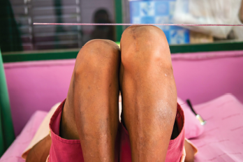
Visual analysis can also be used and is the easiest way to spot a discrepancy. The physician should start by having the patient lie on the exam table, ensuring the patient’s pelvis is flat, and then putting the patient’s legs together. By placing the malleoli of each foot or each leg next to the other, the provider can see whether there appears to be a difference or not.
Manual measurements can’t discern between functional or structural discrepancies, and they tend to be inexact, depending on the provider’s technique and experience.1,4 Accuracy can also be affected by other issues the patient has. Genu valgum deformity of the knee, for example, causes differences in the length of the leg and progression of OA.5

Dr. Lane
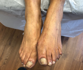
Foot height discrepancy can be a significant contributor to LLD, says George Lane, DPM, a podiatrist at Achilles Foot & Ankle Center, Richmond, Va., who developed a novel technique for evaluating foot contribution to LLD.6
“A mild to moderate foot height discrepancy, which is often just a few millimeters, means there are typically significant misalignments within the joints of at least one of the feet,” Dr. Lane explains. “This can not only have a profound damaging effect on the joints of the feet, but also on ligaments, tendons and joints of the lower extremities and back.”

Dr. Smith
In addition to looking at limb length, evaluating movement and flexibility is an important element of the physical exam, says Craig A. Smith, PT, DPT, Smith Performance Center, Tucson, Ariz. An abnormal gait pattern can be caused by pain, joint effusion or quadriceps weakness, he says, and it’s important to differentiate between these factors before treating.
“If there are joint limitations, you can get what will look like a leg length discrepancy that’s not real,” Dr. Smith says. “That’s a perfect opportunity to try to get the motion back,” potentially by referring the patient to a physical therapist or orthopedist, he says. “But if [the patient has] a true leg length discrepancy and they also have something that’s painful occurring, my recommendation is to … do a gait analysis.”
Dr. Smith looks at five distinct elements during a gait analysis, which helps him identify the underlying issue. For example, a patient with joint effusion doesn’t go through the “normal flexion–extension cycles that occur during the gait cycle,” he says, and the
patient will often “present in the gait analysis like they have a functionally longer leg on one side because they won’t be fully changing their knee like they normally do.” Especially if the patient has been experiencing pain for a long time, they may be compensating for swelling that effectively shut off their quadriceps, so it looks like they have one leg that is long and stiff because they fully extend their knee joint when their heel hits the ground. Alternately, the leg may look functionally shorter if the patient is unable to fully extend their knee because of pain.
In addition to physical examinations, confirming LLD using radiographic imaging is considered the gold standard.1 Other technologies, including ultrasound, laser-
ultrasound and 3-D imaging, may also be used for precision.7–9
Prognosis & Treatment

A visual examination in the physician’s office demonstrates a leg length discrepancy in a patient with unilateral osteoarthritis.
Interventions used to treat LLD vary, depending on the patient and the severity of the discrepancy, and can range from no intervention all the way to surgery. For patients who are experiencing unilateral OA pain related to a clinically significant discrepancy in leg length, treatments may include pharmaceuticals for pain management, physical therapy to address muscle weakness or abnormal gait, or shoe inserts or lifts. By detecting the discrepancy and treating it, further damage to the joint may be prevented.
Lift therapy should be implemented gradually and should be used more conservatively for older patients.4 “If someone has 5 mm or 10 mm of leg length inequality, then generally we would bolster the shorter side with a shoe lift,” Dr. Segal says. He notes that it’s important not to treat more than half the difference—for example, if a patient has an 8 mm difference, the lift would correct 4 mm of it. This is to avoid additional issues such as low-back pain.
There are many options for a rheumatologist to consider when diagnosing & treating a patient with unilateral hip or knee OA. Considering the potential for a discrepancy in leg length & how (& whether) to treat it may be part of
that process.
“I think it is under-appreciated how much the foot may contribute to functional limb-length discrepancies, and the resultant pain and potential arthritic changes that can be associated with this,” Dr. Lane says. “Properly made custom orthotics can play a pivotal role to dramatically reduce or eliminate pain, progression of deformity and joint damage in this situation.”
There may also be reason not to go straight to treating LLD because it can have unintended consequences. For example, prescribing a prosthetic such as a lift or an insert may lead the patient to feel “disabled” or like they can never walk without their shoes on, Dr. Smith says. Instead, physical activity and exercise can be effective ways to treat OA pain and help the patient return to more normal function.10
Recent onset of unilateral knee pain is clinically important, too, and is best treated conservatively to start, Dr. Smith says. “Even if it seems like it’s bone on bone, if it’s only recently painful, you can definitely treat it conservatively, especially if you can normalize the quad. So if you have atrophy of the quadriceps muscle, you get the quad back. If you have joint effusion, you can reduce joint effusion. And if you have significant motion loss, if you can normalize motion and get back to normal gait, actually the prognosis is excellent.”
There are many options for a rheumatologist to consider when diagnosing and treating a patient with unilateral hip or knee OA. Considering the potential for a discrepancy in leg length and how (and whether) to treat it may be part of that process. The good news is that with the proper diagnosis and treatment regimen, which may include working with other healthcare providers, the patient can overcome their pain and get back to living their life.
Abdollah Shams-Pirzadeh, MD, PA, FACR, is a practicing rheumatologist at the AOP Center (Arthritis, Osteoporosis and Pain Management Center), Baltimore.
Kimberly Retzlaff is a freelance medical journalist based in Denver.
References
- Murray KJ, Azari MF. Leg length discrepancy and osteoarthritis in the knee, hip and lumbar spine. J Can Chiropr Assoc. 2015 Sep;59(3):226–237.
- Golightly YM, Allen KD, Helmick CG, Renner JB, Jordan JM. Symptoms of the knee and hip in individuals with and without limb length inequality. Osteoarthr Cartilage. 2009 May;17(5):596–600.
- Harvey WF, Yang M, Cooke TDV, et al. Association of leg-length inequality with knee osteoarthritis: A cohort study. Ann Intern Med. 2010 Mar;152(5):287–295.
- Brady RJ, Dean JB, Skinner TM, Gross MT. Limb length inequality: Clinical implications for assessment and intervention. J Orthop Sports Phys Ther. 2003 May;33(5):221–234.
- Felson DT, Niu J, Gross KD, et al. Valgus malalignment is a risk factor for lateral knee osteoarthritis incidence and progression: Findings from the Multicenter Osteoarthritis Study and the Osteoarthritis Initiative. Arthritis Rheum. 2013 Feb;65(2):355–362.
- Lane G. A novel technique to determine foot contribution to limb-length discrepancy. J Am Podiatr Med Assoc. 2017 Jul;107(4):340–341.
- Krettek C, Koch T, Henzler D, Blauth M, Hoffmann R. [A new procedure for determining leg length and leg length inequality using ultrasound. II: Comparison of ultrasound, teleradiography and 2 clinical procedures in 50 patients.] [Article in German] Unfallchirurg. 1996 Jan;99(1):43–51.
- Rannisto S, Paalanne N, Rannisto PH, Haapanen A, Oksaoja S, Uitti J, et al. Measurement of leg-length discrepancy using laser-based ultrasound method. Acta Radiologica. 2011;52(10):1143–1146.
- Guggenberger R, Pfirrmann CWA, Koch PP, Buck FM. Assessment of lower limb length and alignment by biplanar linear radiography: Comparison with supine CT and upright full-length radiography. AJR Am J Roentgenol. 2014 Feb;202(2):W161–W167.
- Gay C, Chabaud A, Guilley E, Coudeyre E. Educating patients about the benefits of physical activity and exercise for their hip and knee osteoarthritis. Systematic literature review. Ann Phys Rehabil Med. 2016 Jun;59(3):174–183.

