Video: Every Case Tells a Story| Webinar: ACR/CHEST ILD Guidelines in Practice

An official publication of the ACR and the ARP serving rheumatologists and rheumatology professionals

  • Conditions
    • Axial Spondyloarthritis
    • Gout and Crystalline Arthritis
    • Myositis
    • Osteoarthritis and Bone Disorders
    • Pain Syndromes
    • Pediatric Conditions
    • Psoriatic Arthritis
    • Rheumatoid Arthritis
    • Sjögren’s Disease
    • Systemic Lupus Erythematosus
    • Systemic Sclerosis
    • Vasculitis
    • Other Rheumatic Conditions
  • FocusRheum
    • ANCA-Associated Vasculitis
    • Axial Spondyloarthritis
    • Gout
    • Psoriatic Arthritis
    • Rheumatoid Arthritis
    • Systemic Lupus Erythematosus
  • Guidance
    • Clinical Criteria/Guidelines
    • Ethics
    • Legal Updates
    • Legislation & Advocacy
    • Meeting Reports
      • ACR Convergence
      • Other ACR meetings
      • EULAR/Other
    • Research Rheum
  • Drug Updates
    • Analgesics
    • Biologics/DMARDs
  • Practice Support
    • Billing/Coding
    • EMRs
    • Facility
    • Insurance
    • QA/QI
    • Technology
    • Workforce
  • Opinion
    • Patient Perspective
    • Profiles
    • Rheuminations
      • Video
    • Speak Out Rheum
  • Career
    • ACR ExamRheum
    • Awards
    • Career Development
  • ACR
    • ACR Home
    • ACR Convergence
    • ACR Guidelines
    • Journals
      • ACR Open Rheumatology
      • Arthritis & Rheumatology
      • Arthritis Care & Research
    • From the College
    • Events/CME
    • President’s Perspective
  • Search

Type I IFNs in Cutaneous Lupus & Dermatomyositis

Katie Robinson  |  Issue: November 2024  |  October 14, 2024

Clinical Case

The clinical case follows a 29-year-old woman presenting with a six-month history of a pruritic rash that began after a viral illness. The patient’s symptoms include fatigue, hair loss and severe scalp pruritus. She was previously diagnosed with eczema, which did not improve on treatment with antihistamines and topical corticosteroids.

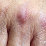
Clinical examination revealed violaceous periorbital edema, scaly red papules on the metacarpophalangeal joints of both hands, dilated capillaries of the proximal nail folds, scaly plaques on both elbows, and excoriated red plaques on her upper chest, back and hips. Antibody testing proved negative. The patient did not report any muscle weakness, strength and creatinine phosphokinase testing appeared normal, and magnetic resonance imaging of the thigh ruled out inflammation or edema.

ad goes here:advert-1
ADVERTISEMENT
SCROLL TO CONTINUE

The patient was diagnosed with clinically amyopathic dermatomyositis and treated with methotrexate, but this did not improve her skin and led to debilitating intractable pruritus. Mycophenolate mofetil treatment provided minimal improvement. However, given the lack of muscle involvement, the patient’s health insurance did not approve using intravenous immunoglobulin (IVIG).

The authors note that patients with clinically amyopathic dermatomyositis comprise up to 60% of dermatomyositis cases in dermatology clinics. As in the clinical case, “skin disease is often refractory to current systemic directed therapies and results in debilitating pruritus and increased morbidity,” the authors write. “Given the effectiveness of IVIG, and the likelihood of success with IVIG in this case, it is essential that dermatomyositis trials include amyopathic dermatomyositis patients so they can access appropriate therapies.”

ad goes here:advert-2
ADVERTISEMENT
SCROLL TO CONTINUE

Cutaneous Lupus Erythematosus vs. Dermatomyositis

Knowledge remains limited regarding both the pathogenesis of dermatomyositis and the drivers of skin disease.

The clinical case “raises several important issues related to the immunologic underpinnings of cutaneous lupus erythematosus and dermatomyositis,” including the roles of type I IFN in triggering skin disease and IFN in the pathogenesis of skin inflammation, the authors write. “Can we apply what we know about IFN-targeted therapeutics in cutaneous lupus erythematosus and dermatomyositis to develop better treatments for skin disease?”

Cutaneous lupus erythematosus and dermatomyositis remain challenging to differentiate, especially if the disease is limited to the skin, with rashes appearing similarly in both conditions. In dermatomyositis, as with cutaneous lupus erythematosus, type I IFNs appear highly upregulated in the blood, muscle and skin, correlating to disease activity. Further, in genetically predisposed individuals, cutaneous lupus erythematosus and dermatomyositis skin are likely induced or triggered by increased IFN exposure, as with sunlight, medications such as immune checkpoint inhibitors and hydroxyurea, immune-stimulating supplements or viral illnesses, including COVID-19.

Page: 1 2 3 | Single Page
Share: 

Filed under:ConditionsMyositis Tagged with:Immunologytype I interferon

Related Articles
    Representation of the molecular structure of interferon a.

    Interferons in Systemic Lupus Erythematosus

    March 21, 2024

    Current knowledge of receptor-ligand interactions, cell signaling, and transcriptional regulation derive from studies of type I interferon. The design of novel therapeutics is informed by the advances in investigation of type I interferon, with the potential for important impacts on patient management.

    Dermatologist, Rheumatologist Discuss Refractory Cutaneous Lupus Case

    September 5, 2022

    As a dermatologist/internist with a career-long subspecialty interest in the cutaneous manifestations of the rheumatic diseases, I found the case of refractory acute cutaneous lupus by Samantha C. Shapiro, MD, in the June 2022 issue of The Rheumatologist intriguing in several ways, and I felt my perspectives on this case might provide additional educational value…

    Interferon Score Predicts AI-CTDs

    November 18, 2018

    People with autoimmune connective tissue diseases produce antibodies against nuclear antigens up to 10 years before they develop clinical features. Anti-nuclear antibodies (ANAs) are also very common, and a small percentage of ANA-positive patients progress to clinical autoimmunity. The question: Is there a reliable way to screen at-risk patients before they develop active autoimmunity and…

    Cancer-Associated Myositis: A Case Report & Review of the Literature

    Cancer-Associated Myositis: A Case Report & Review of the Literature

    February 17, 2019

    Since it was first reported in 1916, a correlation between inflammatory myopathies and cancer has been noted in several studies. Population studies have confirmed this relationship, and the phrase cancer-associated myopathy has entered the vernacular. Over the past decade, research efforts have shifted toward revealing associations between autoantibodies and clinical phenotypes. One subset of auto-antigens…

  • About Us
  • Meet the Editors
  • Issue Archives
  • Contribute
  • Advertise
  • Contact Us
  • Copyright © 2025 by John Wiley & Sons, Inc. All rights reserved, including rights for text and data mining and training of artificial technologies or similar technologies. ISSN 1931-3268 (print). ISSN 1931-3209 (online).
  • DEI Statement
  • Privacy Policy
  • Terms of Use
  • Cookie Preferences