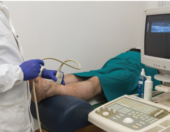
A new study shows that ultrasound can be useful in discriminating gout from non-gout.
Red On/shutterstock.com
The presence of synovial monosodium urate monohydrate (MSU) crystals is the gold standard for diagnosing gout. But a new study, funded in part by the ACR and led by rheumatologists, including Alexis Ogdie, MD, MSCE, evaluated the effectiveness of ultrasound in diagnosing it. The study found that ultrasound can be useful in discriminating gout from non-gout.
“The idea was that if you are a primary care physician or you’re a rheumatologist but can’t do arthrocentesis, what’s the best way to diagnose gout?” says Dr. Ogdie, an assistant professor of medicine and epidemiology at the Hospital of the University of Pennsylvania. “A lot of rheumatologists now have ultrasound as part of their practice, and we wanted to know: What is the sensitivity and the specificity of ultrasound features for the diagnosis of gout?”
In the study, published in February in Arthritis & Rheumatology, the researchers wanted to reflect real-life clinical practice.1 Previous studies had relied on data from relatively small centers of gout expertise, among patients with a long history of the disease and showed that gout can be diagnosed by ultrasound upon detecting hyperechoic articular surface features on the hyaline cartilage (double contour sign, or DCS); a snowstorm appearance suggestive of floating, hyperechoic MSU crystals; or hyperechoic aggregates within the joint or along the tendons, which could indicate tophi.
But the research team, in addition to wanting to capture the capacity of more typical primary care or rheumatology practices to use ultrasound for gout diagnosis, also wanted to better understand the ultrasound features of early gout compared with longstanding disease and of cases with clinically detected tophus and those without.
“Sometimes, a patient doesn’t want arthrocentesis, or there’s not enough fluid in the joint, or they don’t have time,” says Dr. Ogdie. “There are a variety of different reasons, but if the primary care provider has access to ultrasound or it’s performed by a radiologist but they’re not comfortable aspirating that joint, what does that result tell you if you see (or don’t see) DCS, for example. What is the likelihood the patient has gout?”
The Study
To determine this, the researchers used data from the Study for Updated Gout Classification Criteria (SUGAR), a large, cross-sectional study involving 25 international centers. In that study, voluntary ultrasound was performed on 842 subjects, including 416 cases and 408 controls. Cases were subjects ultimately confirmed to have a gout diagnosis, determined by the presence of MSU crystals in fluid aspirates, with swelling in at least one joint or the presence of a subcutaneous nodule. Controls were subjects with gout-like symptoms, including joint swelling, but were negative for gout when tested for MSU crystals.


