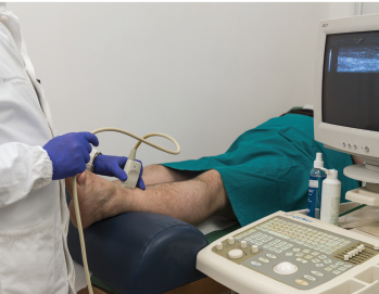
A new study shows that ultrasound can be useful in discriminating gout from non-gout.
Red On/shutterstock.com
The presence of synovial monosodium urate monohydrate (MSU) crystals is the gold standard for diagnosing gout. But a new study, funded in part by the ACR and led by rheumatologists, including Alexis Ogdie, MD, MSCE, evaluated the effectiveness of ultrasound in diagnosing it. The study found that ultrasound can be useful in discriminating gout from non-gout.
“The idea was that if you are a primary care physician or you’re a rheumatologist but can’t do arthrocentesis, what’s the best way to diagnose gout?” says Dr. Ogdie, an assistant professor of medicine and epidemiology at the Hospital of the University of Pennsylvania. “A lot of rheumatologists now have ultrasound as part of their practice, and we wanted to know: What is the sensitivity and the specificity of ultrasound features for the diagnosis of gout?”
In the study, published in February in Arthritis & Rheumatology, the researchers wanted to reflect real-life clinical practice.1 Previous studies had relied on data from relatively small centers of gout expertise, among patients with a long history of the disease and showed that gout can be diagnosed by ultrasound upon detecting hyperechoic articular surface features on the hyaline cartilage (double contour sign, or DCS); a snowstorm appearance suggestive of floating, hyperechoic MSU crystals; or hyperechoic aggregates within the joint or along the tendons, which could indicate tophi.
But the research team, in addition to wanting to capture the capacity of more typical primary care or rheumatology practices to use ultrasound for gout diagnosis, also wanted to better understand the ultrasound features of early gout compared with longstanding disease and of cases with clinically detected tophus and those without.
“Sometimes, a patient doesn’t want arthrocentesis, or there’s not enough fluid in the joint, or they don’t have time,” says Dr. Ogdie. “There are a variety of different reasons, but if the primary care provider has access to ultrasound or it’s performed by a radiologist but they’re not comfortable aspirating that joint, what does that result tell you if you see (or don’t see) DCS, for example. What is the likelihood the patient has gout?”
The Study
To determine this, the researchers used data from the Study for Updated Gout Classification Criteria (SUGAR), a large, cross-sectional study involving 25 international centers. In that study, voluntary ultrasound was performed on 842 subjects, including 416 cases and 408 controls. Cases were subjects ultimately confirmed to have a gout diagnosis, determined by the presence of MSU crystals in fluid aspirates, with swelling in at least one joint or the presence of a subcutaneous nodule. Controls were subjects with gout-like symptoms, including joint swelling, but were negative for gout when tested for MSU crystals.
Trained observers performed the crystal identification. The observers were required to pass a certification test involving a Web-based evaluation and the examination of five vials of synovial fluid.
Ultrasound was also performed on at least one clinically affected joint among SUGAR study subjects. The tests were conducted by ultrasound-trained rheumatologists or radiologists blind to the results of the crystal tests. A hyperechoic band on the surface of the articular cartilage defined double contour sign; tophus was indicated by ultrasound as a hyperechoic, heterogenous lesion surrounded by an anechoic rim; and snowstorm was defined as joint effusion with a snowstorm appearance.
Additionally, the SUGAR study involved gathering subject age, sex, ethnicity, number of joint-swelling episodes, whether or not they utilized urate-lowering therapy, the number of weeks since their last episode, disease duration and whether they had involvement in their metatarsophalangeal joint (podagra). Each subject had a physical examination and the numbers and locations of swollen joints, as well as any suspected tophi, were recorded. Finally, serum urate levels—current and highest recorded in available records—and the presence of any radiographic abnormalities were recorded.
In analyzing the data, the study team examined the sensitivity, specificity, positive predictive value (PPV) and negative predictive value (NPV) of each diagnostic ultrasound feature. They looked at the presence vs. the absence of any one ultrasound feature in cases vs. controls (those with gout and those without). Among these, they examined early disease (fewer than two years since disease onset) vs. late disease and those with and those without clinically suspected tophus.
The researchers also looked at the test characteristics of having any one, two or three ultrasound features. To understand any factors that may influence the results, they also performed statistical analysis among cases to examine any other demographic or clinical associations with having a positive test. They performed additional sensitivity analysis, including whether a subject with a positive test also had a tender or swollen first MTP joint.
They found that sensitivity (the percentage of positive ultrasound results among those positive for gout) for DCS was 60.1%, specificity (the percentage of negative ultrasound results among those negative for gout) was 91.4%, PPV (the likelihood someone with a positive ultrasound result had gout) was 87.7% and NPV (the likelihood that someone with a negative ultrasound did not have the disease) was 69.3%. For tophus identified by ultrasound, sensitivity was 46%, specificity was 94.9%, PPV was 90%, and NPV was 65.6%. For snowstorm, sensitivity was 30.3%, specificity was 90.9%, PPV was 77.2%, and NPV was 56.3%.
When any one feature was considered a positive ultrasound for gout, sensitivity was 76.9%, specificity was 84.3%, PPV was 83.3%, and NPV was 78.2%.
Comparing early to late disease, the researchers found no significant difference in sensitivity or specificity. Subjects with tophi on clinical examination had higher sensitivity and PPV compared with those without clinically detected tophi, while specificity and NPV were lower.
“Early gout didn’t look that different from late gout,” says Dr. Ogdie.
Subjects with an actively tender or swollen first MTP joint and those with elevated serum urate levels had lower specificity and NPV for all ultrasound features.
The researchers also found that a positive ultrasound finding for gout was associated with suspected tophi upon examination, higher serum urate level and abnormal radiographic findings, which means that ultrasound may not be necessary to diagnose gout in these patients, but it can still be helpful in patients without obvious gout upon examination.
Conclusions
In general, “The specificity is high,” Dr. Ogdie says, “so we can be fairly confident that a positive ultrasound is suggestive of gout. If it’s negative, we could still be missing people who do have gout.”
In previous studies, gout has been shown to be difficult to discern from calcium pyrophosphate deposition disease (CPDD) by ultrasound, so the study team examined the specificity of ultrasound features for gout among patients with CPDD (confirmed by aspirates). They found sensitivity and PPV were still high for gout.
“Ultrasound is a pretty good test for gout,” Dr. Ogdie says. “Just be aware that it could miss people with gout who have a negative test.”
Kelly April Tyrrell writes about health, science and health policy. She lives in Madison, Wis.
Reference
- Ogdie A, Taylor WJ, Neogi T, et al. Performance of ultrasound in the diagnosis of gout in a multicenter study comparison with monosodium urate monohydrate crystal analysis as the gold standard. Arthritis Rheumatol. 2017 Feb;69(2):429–438.


