 From the first substantial argument in the 19th century that uric acid played a role in gout, it took about 100 years for the medical community to accept its role in triggering acute inflammatory gout attacks. Two papers, both published in 1962, helped demonstrate the link between uric acid and acute gout attacks, quickly opening the way for successful treatment with urate-lowering therapies.1,2
Historical Background
Known to the Egyptians and the Greeks, gouty arthritis was one of the first diseases to be recognized as a distinct clinical entity. Dutch pioneer of microscopy Antonie van Leeuwenhoek was the first to describe the appearance of crystals from a gouty tophus in 1679, although the chemical composition was then unknown.3 In 1797, the English chemist William Hyde Wollaston, MD, demonstrated the presence of uric acid in gouty tophi, providing some of the earliest evidence of a possible pathophysiologic connection between high uric acid levels and gout.1
Garrod’s Work
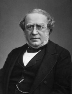
In 1854, English physician Alfred Baring Garrod, MD, described the famous thread test that he had developed—a semi-quantitative method for measuring the uric acid in blood or urine. Using it, he demonstrated that most of his gouty patients were hyperuricemic.1 Through research and his extensive clinical experience, Dr. Garrod came to believe that deposited urate crystals triggered the inflammation response in gouty inflammation.

Dr. Garrod
Dr. Garrod also carefully distinguished the characteristics of gout from rheumatoid arthritis—then termed rheumatism—arguing the two had distinct clinical presentations and underlying disease processes that did not morph into one another. In his mammoth 1876 treatise on the subject, Dr. Garrod noted the following as part of his key characterizations of gout:
Uric acid … is invariably present in the blood in abnormal quantities. … True gouty inflammation is always accompanied by a deposition of urate of soda in the inflamed part. … The deposit is crystalline and interstitial. … The deposited urate of soda may be looked upon as the cause, not the effect of the gouty inflammation. … In no disease but true gout is there a deposition of urate of soda in the inflamed tissues.4
However, many did not accept this explanation, and the clinicians of the time continued to debate gout’s cause. In 1899, a young Swiss internist, Max Freudweiler, MD, outlined the debate of the time. He noted that Garrod’s followers believed the oversaturation of body fluids led to the deposition of crystalline uric acid salts into the tissues.
“This school believes that crystal deposition is the primary step in the development of gouty tophi and therefore attribute tissue necrosis to the damaging effect of the crystals,” he said. “In opposition are other authors … who defend the idea that the tissue necrosis of the gouty lesion is the primary event, and the deposition of uric acid salts is secondary to this.”5
In his own work injecting urate crystals into rabbits, Dr. Freudweiler observed evidence of acute inflammation and tissue necrosis in those injected.5 Some other early research
also found inflammatory reactions resulting from the injection of crystalline uric acids or other forms of urates into living tissue.5 But much of this work fell into obscurity, and Dr. Garrod’s work was left unconfirmed.
Intervening Years
In the following years, the medical community did come to accept a pathophysiologic role between tophaceous gout and hyperuricemia. Salicylates, the first uricosuric agents when given in high doses, and then later agents, such as probenecid, could successfully lower serum urate and gradually dissolve tophaceous gout deposits.2,3
Through the 1950s, however, medical texts were noncommittal about the relationship between urates and the pathogenesis of acute inflammatory gouty attacks, and the mechanism of acute gouty arthritis was studied relatively little.2
It was well established by then that some, but not all, people with hyperuricemia suffered attacks of acute gouty arthritis. On the other hand, when solutions of non-crystallized sodium urate were injected into humans, this had not seemed to trigger a gout flare, nor did the administration of large doses of uric acid delivered intravenously.1 At the time, this was a primary point made by those arguing that urates played no role in triggering acute gouty flares.2
Modern Study of Gout
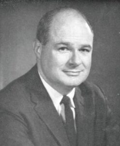
One might argue that the modern study of gout began in 1961, with work led by Daniel J. McCarty Jr., MD, then head of rheumatology at the University of Pennsylvania, Philadelphia.6,7 Using polarized light microscopy, McCarty et al. were able to show crystals of monosodium urate in gouty synovial fluid, often in the process of undergoing phagocytosis. These had been much more difficult to see via the standard light microscopy used previously.

Dr. McCarty
The next year, two key complementary papers in the Journal of the American Medical Association (JAMA) and The Lancet helped definitively establish the role of elevated serum urate and sodium urate crystals in triggering acute gout flares.1,2
The Lancet Article
Dr. McCarty followed up on his important findings in uric acid crystal imaging with his trainee, James S. Faires, MD. Using themselves as the subjects, the two injected uric acid crystals (purified from a gouty tophus and put in solution) into one knee. They injected saline solution without urate crystals into their other knees as a control.1
A few hours later, both experienced excruciating inflammatory gout-like sensations in the knees injected with uric acid crystals. Although they had not initially planned on it, the two ultimately opted to take pain medications and hydrocortisone due to the severity of their symptoms. Follow-up experiments in dogs showed a similar effect.1
The authors noted, “This preliminary work indicates that sodium urate in crystalline form is probably important in the production of acute gouty arthritis. The exact mechanism response for the intense inflammatory arthritis is unknown … The pathogenesis of the acute gouty attack seems to be related to the deposition of sodium-urate crystals in the synovial fluid.”1
The authors also pointed out key questions raised by their work, such as: 1) Why are certain individuals with high uric acid blood levels able to keep their urate in solution? 2) Why are certain tissues in the body more susceptible to deposition? 3) What are the factors responsible for the intense response to sodium urate crystals in synovial fluid?1
JAMA Article
J. Edwin Seegmiller, MD, and his colleagues at the National Institute of Arthritis and Metabolic Diseases of the Public Health Service (est. 1950; later the National Institute of Arthritis and Musculoskeletal and Skin Diseases), Bethesda, Md., published an important, complementary article in JAMA that same year.2
Most previous related work showing an inert inflammatory response to injections had not used crystallized forms of solid sodium urate. In contrast, Dr. Seegmiller and his team injected microcrystalline particulate forms of solid sodium urate into the knees of 12 patient volunteers who had previously had gout disease flares. Each was also injected with an amorphous, non-crystalline solution of sodium urate into the other knee.2
Within a couple of hours, all patients showed signs of pain, warmth and/or effusion in the joints injected with the microcrystalline particulate forms of sodium urate. Several patients described their symptoms as similar to those of a spontaneous gouty attack. Aspirated synovial fluid from the joints was examined under polarized microscopy. This revealed leukocytosis and extensive phagocytosis of the urate crystals.2
In contrast, injection with the amorphous, non-crystallized solution of sodium urate produced very little inflammatory response, one totally absent in the majority of participants. This was consistent with earlier reports from the 1920s, which had failed to produce an inflammatory reaction using a similar preparation.2
“The detailed sequence of events that occurs in the pathogenesis of acute gouty arthritis is yet to be demonstrated experimentally,” the authors noted. “We would propose that in order for acute gouty arthritis to develop, the following conditions must be met: 1) Needle-like crystals of sodium urate must be present. 2) An inflammatory reaction against sodium urate crystals must be elicited.”2
These two papers and other closely related work at the time helped change the paradigm of gout treatment relatively quickly.1,2 Clinical evidence followed rapidly, which supported allopurinol’s use in gout as a urate-lowering therapy.8 The drug was approved for use in the U.S. in 1966, after which it soon became widely prescribed. And in the ACR’s most recent 2020 gout guideline, it is still a cornerstone of preventive gout treatment.9
Present Day
In the intervening years, we have learned quite a bit about specifically how urate crystals help initiate and sustain gouty inflammation through stimulating cellular inflammatory responses and how they also can contribute to long-term inflammation and chronic gouty synovitis. We’ve also learned about some of the local factors hypothesized to play a role in the crystallization of monosodium urate from the blood.10 But many questions remain.
Ted R. Mikuls, MD, MSPH, the Stokes-Shackleford Professor of Rheumatology and vice chair for research at the University of Nebraska Medical Center, Omaha, was one of the authors of the “2020 American College of Rheumatology Guideline for the Management of Gout.”9

Dr. Mikuls
“Questions that were raised in these articles [from 1962] are still being addressed in our gout guideline, which is kind of amazing,” he notes. “To his credit, Garrod was already saying uric acid was a risk factor for gout flares 100 years before these articles were published in the early 1960s. Despite this, the precise links between hyperuricemia and gout flare are still not completely known, but perhaps history tells us not knowing something 60 years later isn’t so bad.”
Seegmiller et al. noted that, at the time, no consistent relationship had been found between the amounts of uric acid in the blood or urine and acute attacks or remissions; this was one of arguments made by some against a role for serum urate in acute gouty arthritis.
“In 60 years, we haven’t fully worked that out,” says Dr. Mikuls. “It’s not a debate about whether it’s the case; it’s a debate about the magnitude, about the relationship of a serum urate level on therapeutics.”
Whether or not to use a treat-to-target strategy for serum urate and the ideal target level have been debated vigorously in recent years. The American College of Physicians’ 2017 guideline did not recommend treating to a specific serum urate target using a serum urate-lowering therapy, such as allopurinol.11 In contrast, the ACR 2020 gout guideline recommends clinicians treat to a target serum urate of 6 mg/dL to reduce the risk of flares, based on some newer evidence.9
“To me,” adds Dr. Mikuls, “that is probably the most important concept in the 2020 ACR gout guideline.”
Another argument against the idea of uric acid’s role in gout flares 60 years ago was the marked hyperuricemia seen in some people without gout. “While we have some more thoughts on that now, the same question was raised in this JAMA article from 60 years ago,” says Dr. Mikuls. “We still don’t really have an answer.”
Others at the time argued that elevated uric acid could not be a potential trigger of gouty attacks, as lowering the serum uric acid by uricosuric drugs did not relieve acute attacks of gout. On the contrary, it was known the administration of these drugs might exacerbate an acute attack.
“By the time gout patients have their first flare, they have built up huge urate stores in their body, and it takes a long time to deplete those,” Dr. Mikuls explains. “That’s one thing we really understand that we probably didn’t 60 years ago, that you have to slowly empty that uric acid load, and that takes—with effective conventional therapy—probably a couple of years for most patients.”
Whether or not to use a treat-to-target strategy for serum urate & the ideal target level have been debated vigorously in recent years.
Similarly, it was known at the time that colchicine, a drug with no effect on uric acid levels, could effectively treat acute gout attacks. This was taken by some as partial evidence that uric acid did not play an important role.
The ACR gout guideline recommends using a prophylactic anti-inflammatory medication, such as colchicine, when first starting patients on urate-lowering medicines, such as allopurinol, to help prevent rebound flares.9 Dr. Mikuls also points out that many patients—and even some medical professionals—get confused about the different roles in the treatment of gout for anti-inflammatory medications vs. urate-lowering therapies, which can negatively influence medication adherence and flare prevention.
Certainly, self-experimentation by physicians, which played a role in many important and historic medical discoveries, especially in the first part of the twentieth century, is now not viewed through the same lens. “I’d like to find the rheumatologists who would be brave enough to repeat these studies on themselves,” says Dr. Mikuls. “Gout is hugely painful. People who get it say, ‘This is the worst thing I’ve ever had.’ To do this to yourself is gutsy and shows a high level of commitment and intellectual curiosity.”
Ruth Jessen Hickman, MD, is a graduate of the Indiana University School of Medicine. She is a freelance medical and science writer living in Bloomington, Ind.
References
- Faires JS, McCarty DJ. Acute arthritis in man and dog after intrasynovial injection of sodium urate crystals. Lancet. 1962 Oct 6;2(7258):682–685.
- Seegmiller JE, Howell R, Malawista SE. The inflammatory reaction to sodium urate: Its possible relationship to the genesis of acute gouty arthritis. JAMA. 1962 May 12;180(6):469–475.
- Nuki G, Simkin PA. A concise history of gout and hyperuricemia and their treatment. Arthritis Res Ther. 2006;8 Suppl 1(Suppl 1):S1.
- Garrod AB. Treatise on Gout and Rheumatic Gout (Rheumatoid Arthritis). 3rd ed. Longmans, Green & Co Inc; 1876.
- Brill JM, McCarty DJ. ‘Studies on the nature of gouty tophi’ by Max Freudweiler, 1899. (An inflammatory response to injected sodium urate.) An abridged translation, with comments. Ann Intern Med. 1964 Mar;60:486–505.
- Malawista SE, de Boisfleury AC, Naccache PH. Inflammatory gout: Observations over a half-century. FASEB J. 2011 Dec;25(12):4073–4078.
- McCarty DJ, Hollander JL. Identification of urate crystals in gouty synovial fluid. Ann Intern Med. 1961 Mar;54:452–460.
- Rundles RW, Wyngaarden JB, Hitchings GH, et al. Effects of a xanthine oxidase inhibitor on thiopurine metabolism, hyperuricemia and gout. Trans Ass Am Physicians. 1963;76:126–140.
- FitzGerald JD, Dalbeth N, Mikuls T, et al. 2020 American College of Rheumatology guideline for the management of gout. Arthritis Care Res (Hoboken). 2020 Jun;72(6):744–760.
- Ragab G, Elshahaly M, Bardin T. Gout: An old disease in new perspective—A review. J Adv Res. 2017 Sep;8(5):495–511.
- Qaseem A, Harris RP, Forciea MA, et al. Management of acute and recurrent gout: A clinical practice guideline from the American College of Physicians. Ann Intern Med. 2017 Jan 3;166(1):58–68.
- Weisse AB. Self-experimentation and its role in medical research. Tex Heart Inst J. 2012;39(1):51–54.

