 EULAR 2021—Temporal arteritis is the term previously applied to giant cell arteritis (GCA), given the predilection for this large vessel vasculitis to involve the temporal arteries. However, recent work on the manifestations of this disease demonstrate GCA is far more complex than previously understood. At EULAR 2021, Sarah Mackie, BMBCh, PhD, MRCP, associate clinical professor in vascular rheumatology, University of Leeds, U.K., provided an overview of the diagnosis and management of GCA.
Evaluation & Diagnosis
Diagnosing GCA can be challenging. Many of the symptoms are systemic and non-specific, and even more specific findings, such as anterior ischemic optic neuropathy, can be seen in other conditions or have close mimics (i.e., non-arteritic anterior ischemic optic neuropathy), noted Dr. Mackie. Temporal artery biopsy is frequently used to provide a pathologic diagnosis, but false-negative biopsy results are not uncommon.
Patients with GCA have traditionally been treated with high-dose glucocorticoids with a prolonged, slow taper, but the relapse rate may be as high as 50%. Risk factors for relapse include elevated baseline inflammatory markers and large vessel involvement.
Vision & GCA
Permanent vision loss is among the most feared complications of GCA. On this subject, Dr. Mackie noted the potential benefits of a fast-track evaluation pathway. Monti et al. conducted a study in which 63 patients with suspected GCA were fast-tracked to evaluation with an expert rheumatologist within one day and underwent clinical evaluation and color duplex sonography. The outcomes for these patients were compared with a historical cohort of 97 patients who were evaluated via the conventional approach prior to introduction of the fast-track assessment process.
The researchers found the relative risk of blindness in the conventional group was 2.11 (95% confidence interval [CI] 1.02–4.36; P=0.04) compared with the fast-track assessment group.1 These results indicate fast-track assessments may provide significant benefits for patients in terms of preventing long-term disability from disease.
With regard to screening for disease, Dr. Mackie discussed the halo sign, a periluminal dark ring detected by color Doppler ultrasonography of the temporal arteries. This sign can be a helpful, non-invasive indicator of potential GCA. However, it’s possible to see the halo sign in the context of conditions other than GCA, such as atherosclerosis, narrow-angle glaucoma, non-Hodgkin’s lymphoma, skull base osteomyelitis and neurosyphilis. Dr. Mackie said some evidence indicates patients with GCA who demonstrate a halo sign around the vertebral artery may be at increased risk of stroke.2
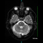
Diplopia in patients with GCA is a concerning indication of possible impending vision loss. Mournet et al. evaluated 14 patients with GCA who presented with diplopia. Using high-resolution, 3D, contrast-enhanced magnetic resonance imaging (MRI), researchers detected third cranial nerve enhancement in seven out of eight patients with third cranial nerve impairment. None of the patients without diplopia showed third cranial nerve enhancement on MRI.
The MRI finding has been called the checkmark sign because that’s the shape formed by the T2-weighted hyperintensity within the orbital apex due to the typical branching of the third cranial nerve within the superior orbital fissure.3 The apparent specificity of this finding for third cranial nerve involvement in GCA may provide another non-invasive way to evaluate for the disease and, in particular, identify patients at risk for ocular ischemia.
Research
Dr. Mackie intimated that many rheumatologists would recall the 2017 publication of the Giant-Cell Arteritis Actemra (GiACTA) trial. In this phase 2, randomized, placebo-controlled study, a significantly larger number of patients with GCA receiving tocilizumab, either every week or every other week, achieved steroid-free remission at 52 weeks than those receiving placebo.4
In a further evaluation of this study, Stone et al. investigated glucocorticoid doses and serologic findings in patients with GCA flares. Although 24% of patients treated with tocilizumab experienced flare, 58% of patients treated with placebo experienced flare. Many of the flares in these groups occurred while patients were still receiving prednisone at doses greater than 10 mg per day.
Another interesting finding was the degree of discordance between the inflammatory marker level and the status of disease activity. Not only did several flares—33 in the tocilizumab-treated group and 20 in the placebo group—occur with patients demonstrating normal C-reactive protein (CRP) levels, but also more than half of placebo-treated patients with elevated CRP levels did not experience flare.5
This study indicates clinicians must carefully monitor for flares of GCA in patients treated with and without tocilizumab. Relying on inflammatory markers alone may not be sufficient to gauge disease activity.
Disease Activity
Dr. Mackie asked: How should rheumatologists best monitor disease activity in patients being treated for GCA? Rheumatologists must take into account several parameters, including signs and symptoms of active disease, which require expertise to assess; blood testing, which is useful, albeit imperfect; and possibly imaging results via ultrasound, magnetic resonance angiogram and FDG-PET/CT, though abnormalities can persist on imaging without disease being clinically active.
Disease activity is important with respect to assessing its impact on the patient’s overall health and life. Early potential complications in GCA include stroke and vision loss. Other manifestations include infections, diabetes, fractures, cardiovascular events and aortic dilatation. There is certainly a life impact on patients in the form of burden of symptoms from GCA and its treatment. Additionally, the total cost to individuals and society in terms of deaths from the disease and healthcare expenses is sizable.
To end her talk, Dr. Mackie stated we must ensure research on GCA accounts for the diversity of patient groups. This includes looking at patients with GCA at either end of the age spectrum, ethnic minorities with GCA and patients in both rural and urban areas who suffer from the disease. Moreover, evaluating how the frequency of follow-up and access to healthcare affects patient outcomes will be essential to helping patients with GCA.
By applying the clinical pearls from this lecture to daily practice, rheumatologists can ensure patients with GCA are diagnosed in a timely manner and given the best possible care.
Jason Liebowitz, MD, completed his fellowship in rheumatology at Johns Hopkins University, Baltimore, where he also earned his medical degree. He is currently in practice with Skylands Medical Group, N.J.
References
- Monti S, Bartoletti A, Bellis E, et al. Fast-track ultrasound clinic for the diagnosis of giant cell arteritis changes the prognosis of the disease but not the risk of future relapse. Front Med (Lausanne). 2020 Dec 8;7:589794.
- Soares C, Costa A, Santos R, et al. Clinical, laboratory and ultrasonographic interrelations in giant cell arteritis. J Stroke Cerebrovasc Dis. 2021 Apr;30(4):105601.
- Mournet S, Sené T, Charbonneau F, et al. High-resolution MRI demonstrates signal abnormalities of the 3rd cranial nerve in giant cell arteritis patients with 3rd cranial nerve impairment. Eur Radiol. 2021 Jan 13. Online ahead of print.
- Stone JH, Klearman M, Collinson N. Trial of tocilizumab in giant cell arteritis. N Engl J Med. 2017 Oct 12;377(15):1494–1495.
- Stone JH, Tuckwell K, Dimonaco S, et al. Glucocorticoid dosages and acute-phase reactant levels at giant cell arteritis flare in a randomized trial of tocilizumab. Arthritis Rheumatol. 2019 Aug;71(8):1329–1338. Epub 2019 Jul 3.

