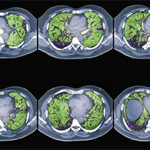
Among surgically biopsied patients with SSc-associated ILD (SSc-ILD), the NSIP pattern is most common, identified in 64–78% of SSc-ILD subjects in various studies.4-6 All of these studies are potentially affected by selection bias. Similarly, in small (also obviously biased) case series, the NSIP pattern predominated among surgically biopsied subjects with PM/DM, primary Sjögren’s, and mixed connective tissue disease. In contrast, although also based on limited data, studies from several investigators suggest that the UIP pattern predominates in RA-associated ILD (RA-ILD).7,8
Results from several studies have shown that, among those with IIP, it is the histologic pattern that drives prognosis. While controlling for other potentially important predictors, patients with UIP pattern have worse survival than patients with NSIP (or any other) pattern lung injury. What is far less clear, and a focus of much debate and ongoing research, is to what degree the histopathologic pattern identified in SLBx specimens from patients with CTD-ILD impacts prognosis. Bouros and colleagues studied 80 subjects with SSc-ILD who had undergone SLBx and found that five-year survival was not significantly different between those with UIP and those with NSIP-pattern histology. Rather, mortality was associated with lower initial diffusing capacity of the lung for carbon monoxide, lower initial forced vital capacity (FVC), and a decline in diffusing capacity over time.4 Similarly, Park and colleagues studied survival in 93 individuals with CTD-ILD who underwent SLBx (37 with SSc-ILD, 28 with RA-ILD, 11 with Sjögren’s-associated ILD, eight with myositis-associated ILD, and nine others).8 They found that age, duration of dyspnea, and FVC were associated with survival, but histological pattern (i.e., NSIP pattern vs. UIP pattern) was not. Results from other studies that included subjects with CTD-ILD suggest the presence of fibrosis (i.e., UIP pattern) is associated with worse survival.9
Because of the conflicting data, many clinicians are uncertain about whether and when to have their patients with CTD-ILD undergo SLBx. On the one hand, many physicians believe that, as is the case with the IIP, UIP-pattern histology confers a worse prognosis than NSIP-pattern histology among patients with CTD-ILD. Physicians holding this view would have their CTD-ILD patients undergo SLBx because the results could better define disease trajectory. On the other hand, some clinicians choose not to send their CTD-ILD patients for SLBx, either because they are not convinced that the histological pattern trumps pulmonary physiology in determining prognosis, or more likely because they don’t expect the results to change management: they plan to treat (or perhaps already are treating) their CTD-ILD patients with immunosuppressive medications, and the pattern identified in SLBx specimens would not affect their choice of therapeutic agent or anticipated duration of therapy. There is greater consensus about the utility of a surgical lung biopsy in patients with CTD-ILD when an etiology for the ILD other than CTD (e.g., hypersensitivity pneumonitis) is seriously considered or when imaging features suggest malignancy or infection (e.g. progressive nodules, cavitation, consolidation, pleural thickening, or effusion).

