Editor’s note: In this recurring feature, we first present a series of images (this page) for your review, and then a brief discussion of the findings and diagnosis (p. 63). Before you turn to the discussion, examine these images carefully and draw your own conclusions. History For the discussion, click here. A 55-year-old man on…
Jennifer L. Demertzis, MD, is an assistant professor of radiology in the musculoskeletal section at the Mallinckrodt Institute of Radiology (MIR) at Washington University School of Medicine in St. Louis, Mo., where she also completed her residency and fellowship training. She is an assistant program director of the MIR radiology residency and directs the resident curriculum for the musculoskeletal section. Since 2010, she has collaborated with the Washington University Department of Rheumatology to present an ongoing series of grand round lectures focusing on the radiologic features of rheumatologic disease in the musculoskeletal system. She practices all aspects of musculoskeletal radiology, with a particular interest in the imaging of inflammatory and infectious disorders and musculoskeletal ultrasound.
Articles by Jennifer L. Demertzis, MD
Diagnostic Imaging in Patient with Left Hip Pain: Findings
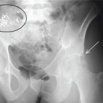
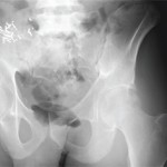
View the question. Findings/Diagnosis The AP radiograph of the left hip (see Figure 1) shows periarticular, well-defined erosions of the left hip (white arrow) without joint space narrowing or osteophytes. There is no fracture. There are surgical clips and a calcified mass in the right hemipelvis (black ellipsis), representing a failed renal transplant. The coronal STIR…
Diagnostic Imaging in Patient with Atraumatic Left Shoulder Pain: History
Editor’s note: In this recurring feature, we first present a series of images (this page) for your review, and then a brief discussion of the findings and diagnosis. Before you turn to the discussion, examine these images carefully and draw your own conclusions. History A 56-year-old man with end-stage renal disease on peritoneal dialysis presents…
Diagnostic Imaging in Patient with Atraumatic Left Shoulder Pain: Findings
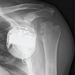
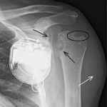
View the question. Findings/Diagnosis The AP radiograph of the left shoulder (see Figure 1) shows erosions of the proximal humeral and glenoid articular surfaces (black arrows) without joint-space narrowing. There is a well-defined marginal erosion with overhanging edge at the junction of the proximal humeral articular surface and rotator cuff insertion on the greater tuberosity (ellipse)….
Diagnostic Imaging in Patient with Soft Tissue Calcification: Findings
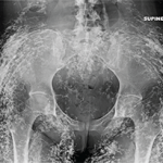
View the question. Findings/Diagnosis The radiographs demonstrate diffuse sheetlike and tumefactive calcifications throughout the subcutaneous tissues, muscle and fascia of the pelvis and right hand. The underlying bones and joint spaces appear normal. The differential diagnosis for soft tissue calcification is extensive and includes metabolic disturbances (particularly of calcium and phosphate), trauma (e.g., injection sites,…
Diagnostic Imaging in Patient with Soft Tissue Calcification: History
Editor’s note: In this recurring feature, we first present a series of images (this page) for your review, and then a brief discussion of the findings and diagnosis. Before you turn to the discussion, examine these images carefully and draw your own conclusions. History These radiographic images are of a 25-year-old woman with a known…
The Diagnostic View: Assess Your Rheumatology Knowledge
Radiograph, MR images of a 55-year-old man with chronic back pain
The Diagnostic View: Our Findings
Radiograph, MR images reveal symmetric erosive sacroiliitis in a patient who was diagnosed with ankylosing spondylitis
The Diagnostic View: Assess Your Rheumatology Knowledge
Radiograph, MR images of 54-year-old woman with right elbow pain
The Diagnostic View: Our Findings
Radiograph, MR images reveal pigmented villonodular synovitis in 54-year-old woman with right elbow pain