Using vibrational spectroscopy, investigators have discovered a characteristic signature in the blood of fibromyalgia patients that is distinct from other clinical conditions, including RA, OA and SLE…

Using vibrational spectroscopy, investigators have discovered a characteristic signature in the blood of fibromyalgia patients that is distinct from other clinical conditions, including RA, OA and SLE…

Throughout their training and practice, physicians become adept at pattern recognition as a means to efficiently connect and synthesize seemingly disparate laboratory, physical exam, and radiologic and historical findings into a coherent theory for what likely ails the patient sitting in front of them. This inductive method of reasoning is necessary because, based on these…
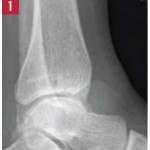
Editor’s note: In this recurring feature, we first present a series of images (this page) for your review, and then a brief discussion of the findings and diagnosis. Before you turn to the discussion, examine these images carefully and draw your own conclusions. History A 49-year-old woman presents with one year of chronic left ankle…
Radiographic imaging showed circumferential soft tissue swelling of the ankle with a soft-tissue density seen in the tibiotalar and posterior subtalar joints, as well as a large, lobulated effusion. MRI of the left ankle shows cystic changes within the talus and first cuneiform bones, as well as a lobulated abnormal soft tissue density with low…
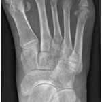
View the question. Findings/Diagnosis An anteroposterior (AP) radiograph of the right foot shows hallux valgus of the first metatarsal phalangeal (MTP) joint, erosive changes at the first and fifth metatarsal bones and degenerative changes at the fourth and fifth metatarsal-cuboid joints. An AP radiograph of the left foot shows extensive erosive and degenerative changes at…

Editor’s note: In this recurring feature, we first present a series of images (this page) for your review, and then a brief discussion of the findings and diagnosis. Before you turn to the discussion, examine these images carefully and draw your own conclusions. History A 33-year-old woman with a 16-year history of systemic lupus erythematosus…
Radiograph, MR images of a 55-year-old man with chronic back pain
Radiograph, MR images reveal symmetric erosive sacroiliitis in a patient who was diagnosed with ankylosing spondylitis
Radiograph, MR images of 54-year-old woman with right elbow pain
The common causes of shoulder pain and diagnostic tests that rheumatologists need to know