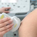
At the 17th Annual Advances in the Diagnosis & Treatment of the Rheumatic Diseases meeting, Dana DiRenzo, MD, MHS, RhMSUS, discussed the use of ultrasound imaging in patients with inflammatory arthritis.
At the 17th Annual Advances in the Diagnosis & Treatment of the Rheumatic Diseases meeting, Dana DiRenzo, MD, MHS, RhMSUS, discussed the use of ultrasound imaging in patients with inflammatory arthritis.
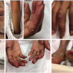
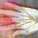
Best Overall: Cutaneous Manifestations of Dermatomyositis These images, submitted by Santhanam Lakshminarayanan, MD, associate professor of medicine in the Division of Rheumatology the University of Connecticut School of Medicine, Farmington, show a 32-year-old Black woman with classic cutaneous manifestations of dermatomyositis: heliotrope rash; periorbital edema with complete closure of the eyes; erythema nodosum on the…

A 27-year-old, left-handed man was referred to our ultrasound clinic for left elbow pain. History The patient had been a pitcher on a Minor League Baseball team. Two years before, he developed sudden, severe medial elbow pain while pitching in a game. The pain was associated with some tingling down the left medial forearm. The…
For most rheumatologists, the key elements of the physical exam—inspection, palpation, percussion and auscultation—have long been second nature, but a fifth modality has grown in importance with respect to making the correct diagnosis: ultrasound. From evaluating for Doppler signal and additional findings indicative of synovitis to identifying bony erosions, chondrocalcinosis, tophi and other articular and…
A 65-year-old woman was referred by an orthopedist to a rheumatologist for left knee pain. Previously, in 2014, she underwent left total knee arthroplasty (TKA) for severe osteoarthritis in a different institution. Following the procedure, she experienced severe chronic anterolateral knee pain at rest, exacerbated by walking. Because she was rendered wheelchair bound and required…
ATLANTA—Two ways to investigate injuries to the upper extremities are by in-depth physical examinations and ultrasound. In a Clinical Practice session at the 2019 ACR/ARP Annual Meeting, Anatomy: Correlating Physical Exam and Ultrasound in Common Sports Injuries of the Upper Extremity, Carlin Senter, MD, FACP, associate professor of primary care sports medicine at the University…
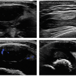
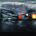
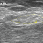
Editor’s note: In this recurring feature, we first present a series of ultrasound images for your review, and then a brief discussion of the findings and diagnosis. Before you scroll to the discussion, examine these images carefully and draw your own conclusions. History A 2-year-old boy with a history of multiple strokes and vertebral artery…
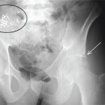
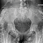
View the question. Findings/Diagnosis The AP radiograph of the left hip (see Figure 1) shows periarticular, well-defined erosions of the left hip (white arrow) without joint space narrowing or osteophytes. There is no fracture. There are surgical clips and a calcified mass in the right hemipelvis (black ellipsis), representing a failed renal transplant. The coronal STIR…
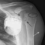
View the question. Findings/Diagnosis The AP radiograph of the left shoulder (see Figure 1) shows erosions of the proximal humeral and glenoid articular surfaces (black arrows) without joint-space narrowing. There is a well-defined marginal erosion with overhanging edge at the junction of the proximal humeral articular surface and rotator cuff insertion on the greater tuberosity (ellipse)….
View the question. Findings/Diagnosis The radiographs demonstrate diffuse sheetlike and tumefactive calcifications throughout the subcutaneous tissues, muscle and fascia of the pelvis and right hand. The underlying bones and joint spaces appear normal. The differential diagnosis for soft tissue calcification is extensive and includes metabolic disturbances (particularly of calcium and phosphate), trauma (e.g., injection sites,…