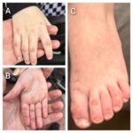
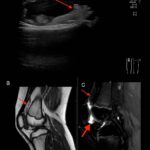
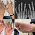
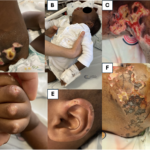
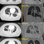
The 2024 Image Competition showcased images representing a diverse range of pediatric patients. Here, we showcase the images from Europe and Central Asia, which were selected by ACR Convergence 2024 attendees in the first plenary session to receive the People’s Choice Award. A Rare Presentation of a Rare Disease An 18-month-old boy from Angola was…
ACR Image Competition 2024 Results, Part 1—Best Overall
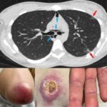
For the 2024 Image Competition, the ACR sought images representing a diverse range of patients with educational or remarkable manifestations of pediatric rheumatic diseases, including autoimmune, inflammatory, infectious and malignant drivers of pediatric rheumatic disease. Here, we showcase the images selected as Best Overall. Cavitary Lung Nodules with Sinus & Skin Disease in Pediatric ANCA-Associated…
ACR Image Competition 2023 Results, Part 5
For the 2023 Image Competition, the ACR sought images representing a diverse range of patients with idiopathic inflammatory myopathies (IIMs) or IIM mimics. Pneumomediastinum & Ulcerative Cutaneous Dermatomyositis These images depict a 22-year-old woman with a recent diagnosis of anti-melanoma differentiation-associated gene 5 (MDA-5) antibody dermatomyositis who sought evaluation of worsening skin rashes and pleuritic…
ACR Image Competition 2023 Results, Part 4
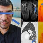
For the 2023 Image Competition, the ACR sought images representing a diverse range of patients with idiopathic inflammatory myopathies (IIMs) or IIM mimics. Unilateral Heliotrope Rash in Anti-MDA-5 Antibody Dermatomyositis These images depict a 36-year-old patient with arthralgias, dyspnea and a unilateral heliotrope rash (image A). A unilateral heliotrope rash is a distinctive, but rare,…
ACR Image Competition 2023 Results, Part 3—People’s Choice Winner
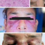
For the 2023 Image Competition, the ACR sought images representing a diverse range of patients with idiopathic inflammatory myopathies (IIMs) or IIM mimics. Chronic Facial Ulcers in Anti-Melanoma-Differentiation-Associated Gene 5 (MDA-5) Antibody Amyopathic Dermatomyositis These images depict a 27-year-old patient who developed erythematous violaceous lesions over his upper chest, face and scalp over six months….
ACR Image Competition 2023 Results, Part 2
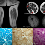
For the 2023 Image Competition, the ACR sought images representing a diverse range of patients with idiopathic inflammatory myopathies (IIMs) or IIM mimics. These images won the Europe and Central Asia region. Glycogen Storage Disease Type V Diagnosed Using Imaging, Biopsy & Genetic Testing These images depict a 55-year-old patient with suspected polymyositis who presented…
ACR Image Competition 2023 Results, Part 1—Best Overall
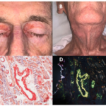
For the 2023 Image Competition, the ACR sought images representing a diverse range of patients with idiopathic inflammatory myopathies (IIMs) or IIM mimics. The images selected as Best Overall by the judges this year come from North America. Systemic Light Chain (AL) Amyloidosis Masquerading as Inflammatory Myositis These images depict an 81-year-old man who presented…
ACR Image Competition 2022 Results, Part 6
Diffuse Cutaneous Scleroderma with Interstitial Lung Disease: Radiological Findings A 44-year-old woman with cutaneous manifestations, tightening of her face with a reduced oral aperture and a healing digital tip ulcer (Figures 1 and 2) has had a known case of diffuse scleroderma since 2008. The patient presented with complaints of shortness of breath along with…
ACR Image Competition 2022 Results, Part 5
Resolution of GAVE After aHSCT for Progressive Systemic Sclerosis A 30-year-old man with RNA polymerase III (Pol III)-positive, diffuse, progressive systemic sclerosis had a persistent microcytic anemia with a hemoglobin level of 8 g/dL and evidence of gastric antral ventral ectasia (GAVE) on gastroscopy. He underwent autologous hematopoietic stem cell transplantation (aHSCT). After six months,…
ACR Image Competition 2022, Part 4
Periungual Erythema & Its Translation on Nailfold Videocapillaroscopy in a Patient with Very Early Systemic Sclerosis A 66-year-old woman presented with Raynaud’s phenomenon and periungual erythema. HEp-2 immunofluorescence assay was positive for antinuclear antibodies, showing a centromere pattern. The presence of anti-centromere antibodies was confirmed by chemiluminescent immunoassay. The patient was diagnosed with very early…