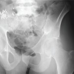
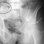
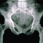
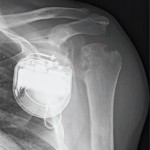
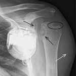
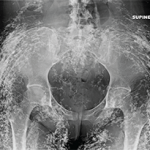
Editor’s note: In this recurring feature, we first present a series of images (this page) for your review, and then a brief discussion of the findings and diagnosis. Before you turn to the discussion, examine these images carefully and draw your own conclusions. History These radiographic images are of a 25-year-old woman with a known…
Diagnostic Imaging in Patient with Elbow Pain: Findings
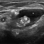
View the question. Transverse (see Figure 1) and longitudinal (see Figure 2) ultrasound images show an anechoic collection (asterisks), which surrounds the distal biceps tendon (b). The collection is compressible (not shown here). The biceps tendon is heterogeneous in echo texture, with fiber dropouts and an irregular border (in the transverse view). The elbow joint…
Imaging in Ankylosing Spondylitis
MRI inflammation, fat and new bone formation in the sacroiliac joints, spine in patients with AS
The Diagnostic View: Assess Your Rheumatology Knowledge
Radiograph, MR images of a 55-year-old man with chronic back pain
The Diagnostic View: Our Findings
Radiograph, MR images reveal symmetric erosive sacroiliitis in a patient who was diagnosed with ankylosing spondylitis
The Diagnostic View: Assess Your Rheumatology Knowledge
Radiograph, MR images of 54-year-old woman with right elbow pain
Coding Corner Answer: October
Coding for facet joint nerve injection for a patient with osteoarthritis
Coding Corner Question: October
Coding for facet joint nerve injection for a patient with osteoarthritis
Choosing Wisely: Recommendations from Medical Specialties Beyond Rheumatology
Practice gems that may help rheumatologists avoid performing costly patient tests, procedures of limited use
EULAR 2014: Ultrasound Use Important for Rheumatology
Experts offer insight into ultrasound for joint counts in rheumatoid arthritis, synovial biopsies, Sjögren’s syndrome
- « Previous Page
- 1
- …
- 3
- 4
- 5
- 6
- 7
- …
- 12
- Next Page »