Highlights from the 2017 EULAR Congress MADRID—Researchers say that whole-body MRI could yield an earlier diagnosis of spondyloarthropathy (SpA) in patients with early inflammatory joint symptoms, according to findings presented in a poster session at the Annual European Congress of Rheumatology (EULAR). The approach could lead to earlier treatment and better outcomes, they say. Investigators…
How to Diagnose Shoulder Pain
WASHINGTON, D.C.—A 70-year-old woman had been diagnosed with rotator cuff disease three years earlier and received an array of treatments. What she hadn’t received was an X-ray. She’d had an MRI, and her doctor—not an orthopedist or a rheumatologist, but a primary care physician—had zeroed in on degenerative changes in her rotator cuff. The problem,…
Lateral Hip Pain: Could It Be Gluteal Tendinopathy?
Researchers recently found that physical tests that involve an active muscle contraction were most useful in identifying gluteal tendinopathy in patients with lateral hip pain. However, patients who were not palpably tender over the greater trochanter were unlikely to have GT detected during an MRI…
How Magnetic Resonance Imaging Technology Can Aid Spondyloarthritis Diagnosis
SAN FRANCISCO—“We haven’t made a lot of progress in ensuring the early diagnosis of spondyloarthritis,” said Walter Maksymowych, MD, FRCP, professor of medicine in the Division of Rheumatology at the University of Alberta and chief medical officer at CaRE (Canadian Research and Education) Arthritis, both in Edmonton. Speaking at the California Rheumatology Alliance 2016 Medical…
Diagnostic Imaging in Patient with Chronic Left Ankle Pain: History
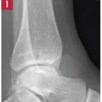
Editor’s note: In this recurring feature, we first present a series of images (this page) for your review, and then a brief discussion of the findings and diagnosis. Before you turn to the discussion, examine these images carefully and draw your own conclusions. History A 49-year-old woman presents with one year of chronic left ankle…
Diagnostic Imaging in Patient with Chronic Left Ankle Pain: Findings
Radiographic imaging showed circumferential soft tissue swelling of the ankle with a soft-tissue density seen in the tibiotalar and posterior subtalar joints, as well as a large, lobulated effusion. MRI of the left ankle shows cystic changes within the talus and first cuneiform bones, as well as a lobulated abnormal soft tissue density with low…
Tofacitinib Decreases Inflammation in Early RA
A recent study used MRI to show that tofacitinib reduces inflammation and inhibition of the progression of structural damage in adults with early RA…
Rheumatology Drug Updates on Brentuximab Vedotin, Tofacitinib Citrate
Brentuximab Vedotin Enters Phase 2 Trials Brentuximab vedotin (Adcetris), an antibody-drug conjugate (ADC) directed at CD30, is currently entering Phase 2 clinical trials for treating systemic lupus erythematosus (SLE).1 The ADC encompasses an anti-CD30 monoclonal antibody, which is attached by a protease-cleavable linker to a microtubule-disrupting agent, known as monomethyl auristatin E (MMAE). The ADC…
EULAR 2015: Imaging in Rheumatology
ROME, Italy—The explosion of imaging technology has made it more important than ever to establish a standardized way in which imaging can and should be used in clinical practice, an expert said in a session at EULAR 2015, the annual congress of the European League Against Rheumatism (EULAR). Marie-Antonietta d’Agostino, MD, PhD, professor of rheumatology…
Diagnostic Imaging in Patient with Left Hip Pain: History
Editor’s note: In this recurring feature, we first present a series of images (this page) for your review, and then a brief discussion of the findings and diagnosis (p. 63). Before you turn to the discussion, examine these images carefully and draw your own conclusions. History For the discussion, click here. A 55-year-old man on…