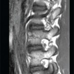
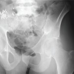
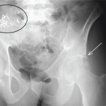
View the question. Findings/Diagnosis The AP radiograph of the left hip (see Figure 1) shows periarticular, well-defined erosions of the left hip (white arrow) without joint space narrowing or osteophytes. There is no fracture. There are surgical clips and a calcified mass in the right hemipelvis (black ellipsis), representing a failed renal transplant. The coronal STIR…
Odds Ratio Explained for MRI Imaging in Ankylosing Spondylitis
In the lead article, “Imaging in Ankylosing Spondylitis” (The Rheumatologist, April 2015), Dr. Pedersen states, “vertebral edges with MRI inflammation were three times as likely to develop new syndesmophytes than vertebral edges without inflammation (odds ratio [OR] 3.3; 95% CI: 1.5–7.4).” This statement is incorrect. An odds ratio of 3 does not imply that an…
Imaging in Ankylosing Spondylitis
MRI inflammation, fat and new bone formation in the sacroiliac joints, spine in patients with AS
fMRI Provides Visual Evidence of Pain
Functional MRI scans that show how pain changes the brain could help rheumatologists develop better approaches to pain relief
EULAR 2014: Cardiac Involvement in Rheumatic Diseases
Experts offer tips for treating, managing cardiac effects; using cardiac magnetic resonance imaging in patients chronic inflammatory conditions
RA Treatment May Be Beneficial Even After Patients Meet Remission Criteria
An analysis of wrist MRIs from participants in the Treatment of Early Aggressive Rheumatoid Arthritis (TEAR) clinical trial indicates that patients continue to show joint inflammation even after two years of early aggressive therapy.
Coding Corner Answer
June’s Coding Answer
Coding MRI Procedures
The Current Procedural Terminology (CPT) manual has six codes to bill for Magnetic Resonance Imaging (MRI) of the upper extremity.
Experts Debate Merits of Peripheral MRI in Diagnosing RA
New technology comes with a host of pros and cons
Envision Arthritis Pathology
MRI advances in RA and OA