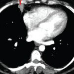
Click here for the case. Discussion Image 1 demonstrates two nodules in the right lung, one over the lateral costophrenic sulcus (2.4 x1.7 cm, not shown); and another in the middle lobe (3.1 x 2.6 cm), with lobulated and spiculated margins (red arrow). There was no lymphadenopathy or pleural effusion. Of note, a normal chest…
The Diagnostic View: Assess Your Rheumatology Knowledge
Editor’s note: In this occasional feature, we first present a series of images (this page) for your review, and then a brief discussion of the findings and diagnosis. Before you read the discussion, examine these images carefully and draw your own conclusions. History A 39-year-old white woman with a three-year history of seropositive rheumatoid arthritis…