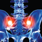
The axial phenotype of psoriatic arthritis (axPsA) is an excellent example of a major controversy in rheumatology that has become the focus of attention because of the emergence of new therapies with different mechanisms of action for alleviating joint inflammation. It was first described in 1961 but, until recently, it has largely remained under the…
Diagnostic Imaging in Patient with Left Hip Pain: History
Editorās note: In this recurring feature, we first present a series of images (this page) for your review, and then a brief discussion of the findings and diagnosis (p. 63). Before you turn to the discussion, examine these images carefully and draw your own conclusions. History For the discussion, click here. A 55-year-old man on…
Diagnostic Imaging in Patient with Left Hip Pain: Findings
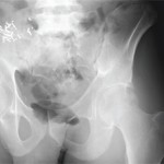
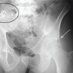
View the question. Findings/Diagnosis The AP radiograph of the left hip (see FigureĀ 1) shows periarticular, well-defined erosions of the left hip (white arrow) without joint space narrowing or osteophytes. There is no fracture. There are surgical clips and a calcified mass in the right hemipelvis (black ellipsis), representing a failed renal transplant. The coronal STIR…
Diagnostic Imaging in Patient with Atraumatic Left Shoulder Pain: History
Editorās note: In this recurring feature, we first present a series of images (this page) for your review, and then a brief discussion of the findings and diagnosis. Before you turn to the discussion, examine these images carefully and draw your own conclusions. History A 56-year-old man with end-stage renal disease on peritoneal dialysis presents…
Diagnostic Imaging in Patient with Atraumatic Left Shoulder Pain: Findings
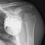
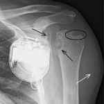
View the question. Findings/Diagnosis The AP radiograph of the left shoulder (see FigureĀ 1) shows erosions of the proximal humeral and glenoid articular surfaces (black arrows) without joint-space narrowing. There is a well-defined marginal erosion with overhanging edge at the junction of the proximal humeral articular surface and rotator cuff insertion on the greater tuberosity (ellipse)….
The Diagnostic View: Assess Your Rheumatology Knowledge
Radiograph, MR images of a 55-year-old man with chronic back pain
The Diagnostic View: Our Findings
Radiograph, MR images reveal symmetric erosive sacroiliitis in a patient who was diagnosed with ankylosing spondylitis
The Diagnostic View: Assess Your Rheumatology Knowledge
Radiograph, MR images of 54-year-old woman with right elbow pain
Physical Therapy Helps Patient with Hip Pain Avoid Surgery
Radiographs reveal greater trochanteric calcific bursitis; guide targeted treatment
Janus Kinase Inhibitors Represent New Tool to Slow Rheumatoid Arthritis
Physicians at the California Rheumatology Alliance 10th Annual Medical & Scientific Meeting in San Francisco report on studies about JAK inhibitors as targeted immunomodulators and disease-modifying therapies in RA