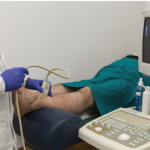
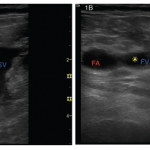
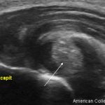
Case A 46-year-old Caucasian female presented to the outpatient rheumatology clinic where she had been followed for several years. Her chief complaint was pain in her right knee, posterior right thigh and right hip that had begun gradually over the previous three weeks. Her past medical history was significant for rheumatoid arthritis (RA), obesity and…
How to Diagnose Shoulder Pain
WASHINGTON, D.C.—A 70-year-old woman had been diagnosed with rotator cuff disease three years earlier and received an array of treatments. What she hadn’t received was an X-ray. She’d had an MRI, and her doctor—not an orthopedist or a rheumatologist, but a primary care physician—had zeroed in on degenerative changes in her rotator cuff. The problem,…
Ultrasound for Underserved Patients
Beverly Johnson, MD, has experienced exactly how rheumatologists and their patients benefit from musculoskeletal ultrasound imaging for the diagnosis and treatment of autoimmune disease…
Rheumatology Coding Corner Answer: Joint Injection with Ultrasound Guidance, No Office Visit
Take the challenge. CPT: 20611-RT, J1040, 89060 ICD-10: M17.11 Coding Rationale Keep in mind, no evaluation and management services are billed because there wasn’t a separate and/or significant reason, other than the knee injection, addressed during the visit. Note: Although the injection was performed via ultrasound guidance, CPT code 76942 should not be billed with…
Rheumatology Coding Corner Question: Joint Injection with Ultrasound Guidance, No Office Visit
A 66-year-old male patient presents to the office with right knee pain. He was in the office two weeks prior for a follow-up visit of his primary osteoarthritis. He received an injection of hyaluronate sodium in his right knee four months before and states that his knee felt like new. He states that everything was…
New Research into Rheumatoid Arthritis, Gout Includes Updates on Methotrexate, Biologics, Ultrasound
LONDON—From optimizing responses to methotrexate, to the efficacy of biologics, to the need for imaging in assessing remission, the literature, as ever, has been lively with explorations of pressing topics in the treatment and management of rheumatoid arthritis. Josef Smolen, MD, chair of rheumatology at the Medical University of Vienna, reviewed many of the highlights…
Ultrasound May Be Useful for Grading Rotator Cuff Tendinopathy
Researchers have developed procedures and assessed their efficacy for the use of ultrasound images to measure the inter-rater reliability of the measurement of structural changes in the tendon of patients with supraspinatus tendinopathy. The standardized procedures proved useful in evaluating patients…
From the Expert: Musculoskeletal Ultrasound Training Benefits Rheumatology Practices & Patients
Eugene Kissin, MD, says the use of musculoskeletal ultrasound can help rheumatologists diagnose and treat disease. Getting the proper training is critical. USSONAR is a training resource that can help…
Diagnostic Imaging in Lupus Patient with Foot Pain: Findings
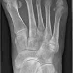
View the question. Findings/Diagnosis An anteroposterior (AP) radiograph of the right foot shows hallux valgus of the first metatarsal phalangeal (MTP) joint, erosive changes at the first and fifth metatarsal bones and degenerative changes at the fourth and fifth metatarsal-cuboid joints. An AP radiograph of the left foot shows extensive erosive and degenerative changes at…
Diagnostic Imaging in Lupus Patient with Foot Pain: History
Editor’s note: In this recurring feature, we first present a series of images (this page) for your review, and then a brief discussion of the findings and diagnosis. Before you turn to the discussion, examine these images carefully and draw your own conclusions. History A 33-year-old woman with a 16-year history of systemic lupus erythematosus…
- « Previous Page
- 1
- 2
- 3
- 4
- 5
- 6
- …
- 8
- Next Page »