Case Presentation
History of present illness
A 66-year-old white woman presented with unexplained, recurrent episodes of high fever, abdominal pain, rash and arthralgias occurring over the previous three years. During typical episodes, the patient experienced flu-like symptoms, followed by fever, abdominal pain and non-bloody diarrhea without tenesmus.
Her temperatures were 101–103ºF, with chills lasting up to 36 hours. She described her abdominal pain as acute and generalized. It was associated with nausea and vomiting, and would often persist for four days with each episode. Episodic, nonpruritic and large erythematous patches over her face and chest could persist up to two weeks.
These episodes were accompanied by diffuse arthralgias, myalgias and swelling in her knees and ankles.
Medical, social & family history
The patient had been diagnosed with fibromyalgia 25 years earlier. She had hypercholesterolemia, hypertension, migraines, rhinitis and asthma. She was taking amitriptyline, aspirin, oxycodone/acetaminophen, pravastatin, sertraline, verapamil, carbamazepine and pantoprazole. She had a remote surgical history of appendectomy, cutaneous basal cell carcinoma and squamous cell carcinoma on her forehead. She also had a history of small meningioma. She did not drink alcohol or smoke. She was allergic to a sulfa antibiotic, which caused rash and laryngeal edema, and to tetracycline, which caused tongue swelling.
Her maternal and paternal families both originated in Czechoslovakia, and there was no known family history of periodic fever syndromes.
Review of systems
Our initial review of systems revealed fatigue, headache, photosensitivity, occasional dry eyes and burning mouth, and low back pain. She had no weight loss, and denied oral ulcers, cough, shortness of breath and chest pain. She also denied abdominal pain, diarrhea, nausea or vomiting.
Physical examination
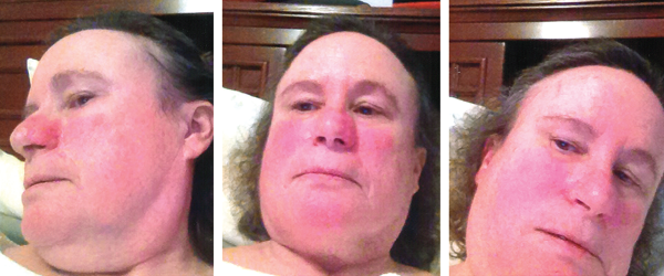
The patient’s vital signs were within normal limits, with a temperature of 97.8ºF. She had no rash, but she brought photographs showing facial erythema (see Figure 1, above). She had no palpable lymphadenopathy, oral ulcers, conjunctivitis or parotid gland enlargement. The patient had chest tenderness, but the cardiopulmonary examination revealed no murmurs, wheezing or rales. On abdominal examination, she had no tenderness, distention, rebound or hepatosplenomegaly. The musculoskeletal examination disclosed no tenderness, redness, warmth or swelling in her joints, and she had no signs of instability of the knees. Her muscle strength was normal, and she had no neurological deficits.

Figure 1: Erythema
Patchy erythema is seen on the cheeks, the bridge of nose, chin and neck.
Laboratory & radiographic evaluation
Complete blood counts, complete metabolic panels and urinalyses were essentially normal on multiple occasions, except for occasional, mild leukocytosis, thrombocytosis and eosinophilia. Her C-reactive protein (CRP) was 12.7 (normal <5 mg/L), and her erythrocyte sedimentation rate (ESR) and serum amyloid A (SAA) were normal.
Skin testing and radioallergosorbent (RAST) assays for both aeroallergens and food hypersensitivities, serum IgE and tryptase level all were negative or normal. Further laboratory tests, including stools for occult blood and lactoferrin, blood cultures, viral hepatitis, parvovirus, Lyme disease, Rickettsia, Babesia, Ehrlichia, Clostridium difficile and Aspergillus all came back negative.
Serologic markers were negative, including autoantibodies directed against nuclear antigens (ANA), extractable nuclear antigens (ENA), double-stranded DNA, neutrophil cytoplasmic antigens (ANCA), and cyclic citrullinated peptides, as well as rheumatoid factor and HLA-B27. Thyroid-stimulating hormone levels were within normal limits, and anti-thyroid peroxidase and thyroglobulin antibodies were negative.
Other tests, such as for creatinine kinase, aldolase, serum protein electrophoresis, uric acid and angiotensin-converting enzymes, were all negative or normal. Serum immunoglobulin quantitation, including IgG subclasses, IgE and IgD levels, was normal, except for IgA at 31 mg/dL (normal 81–463 mg/dL). Tissue transglutaminase IgA antibodies were negative. Genetic testing for celiac disease was negative for the HLA-DQ2/DQ8 haplotypes. Plasma cytokine (IL-1, IL-6 and TNFα) levels were normal.
A chest radiograph was unremarkable, but computerized tomography (CT) of the chest showed a small pericardial effusion. A subsequent echocardiogram was normal. Ultrasound examination of the abdomen was unremarkable; but CT scans of the abdomen and pelvis taken during attacks revealed mesenteric panniculitis (see Figure 2, opposite).
Esophagogastroduodenoscopy showed gastritis, and a colonoscopy was normal except for the presence of benign polyps.
A CT of the patient’s head revealed a left frontoparietal meningioma 0.9×0.7×1.1 cm, and a magnetic resonance angiography (MRA) of her brain was unremarkable.
A radiograph of the knees showed minimal degenerative disease in the left knee. A bone scan did not demonstrate evidence of malignancy, but showed a slight uptake in her carpi and feet.
Next-generation sequencing for periodic fever syndrome in a six-gene panel (MEFV, NLRP3, TNFRSF1A, MVK, NLRP12 and NOD2) was remarkable only for the nucleotide oligomerization domain, containing two (NOD2) variants: c.2798+158 C>T (IVS8+158) and c.2104C>T (R702W).

