Individuals with an excess of nutrition and an excess of soluble urate are often diagnosed with gout. They are diagnosed when their excess urate forms monosodium urate (MSU) crystals that deposit in their articular and soft tissue. The deposits can remain quiescent or trigger the inflammation that is characteristic of gout. Scientists still don’t fully understand why a quiescent urate crystal deposit turns gouty. However, several factors have been identified as playing a role in the pathology of gout. These include the physical characteristics of the MSU crystals, stability of the deposit and systemic triggers from outside the joint. The formed gouty tophi have been characterized, and include not only the MSU crystal deposits, but also macrophages, mast cells, B cells, T cells and other plasma cells.
Most patients with gout have excess body weight, and many have been diagnosed with metabolic syndrome. Scientists have learned that the inflammation that is characteristic of gout is primarily mediated by complement and macrophages, and includes the expression of nuclear factor (NF)-ҡB-dependent proinflammatory cytokines, such as pro-interleukin (IL) 1β, neutrophil chemotactic chemokines CXCL8 (IL-8) and CXCL1. The inflammatory response also centers on activation of the NLRP3 inflammasome and consequent release of IL-1β.
Metabolic Biosensor Enters the Story
A new study suggests the nutritional biosensor AMP-activated protein kinase (AMPK) is the link between nutritional excess and the inflammation that leads to gout.1 AMPK is widely expressed throughout the body and is a driver of stamina. It is activated by stressors that increase the AMP:ATP ratio, such as nutrient deprivation, hypoxia and exercise. As would be expected, individuals who are more physically fit have higher AMPK activity. Previous studies have also shown that excesses in soluble urate suppress tissue AMPK activity. Thus, AMPK has been loosely connected to gout pathophysiology.
Patients with gout know that nutritional excesses and alcohol excesses can trigger gout flares. Metabolic syndrome, obesity, alcohol and multiple nutritional stressors also decrease tissue AMPK activity. In contrast, lifestyle changes, as well as drugs, can activate AMPK. New research adds colchicine to the list of indirect AMPK-promoting drugs. The list includes methotrexate, high-dose aspirin and metformin.
AMPK may thus represent a target for improving efficacy of prophylaxis and treatment of gouty inflammation. Patients may, therefore, be able to improve their gout management by activating their AMPK through exercise and calorie restriction. This new way of thinking about gout expands patient management options beyond the specific dietary restrictions that are designed to decrease the formation of MSU crystals.
Colchicine Drives up AMPK Activity
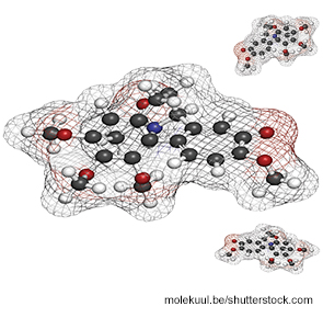
Colchicine is frequently prescribed to patients with gout. It is also known to affect AMPK by inhibiting microtubule polymerization. Colchicine regulates LKB1 activity in macrophages and research has demonstrated that most of its antiinflammatory effects are dependent on the presence of LKB1. The latest research suggests, however, that colchicine is an effective treatment for gout because it activates the nutritional biosensor AMPK.
