Defined by the presence of antiphospholipid antibodies (aPL) in individuals with clinical evidence of thrombosis or pregnancy morbidity, antiphospholipid syndrome (APS) is a systemic autoimmune thrombophilia. Clinical thrombosis, which should be confirmed by objective validated criteria (e.g., imaging studies or histopathology), can occur in the arterial, venous or small vessel vasculature and is not limited to any specific tissue or organ. Pregnancy morbidity is characterized by either fetal loss after the 10th week of gestation, frequent spontaneous abortions before the 10th week of gestation or miscarriage of a neonate due to eclampsia, preeclampsia or placental insufficiency.
Despite the straightforward Sapporo criteria established by Wilson et al. in 1999 and revised in 2006 by Miyakis et al., the clinical spectrum of APS is heterogeneous and can include hematologic, renal, cardiac, dermatologic and neurologic manifestations.1,2 Diagnosis can prove challenging, considering the wide differential and evaluation required to exclude conditions with similar presentations.
Here, we report on a case of APS presenting as digital limb ischemia and gangrene in a patient with a long history of tobacco abuse. We discuss the diagnosis of APS and review pertinent differentials. We also highlight the key features of APS not included in its classification criteria—specifically, the prevalence of cutaneous manifestations—and review the relevant literature.
Case Presentation
A previously healthy 44-year-old man with a 15 pack-year smoking history and who resides in the northeastern U.S. presented with six weeks of fever, polyarthritis and progressive, painful acrocyanosis of his toes and fingers.
His symptoms, swelling and pain in his hands, wrists, ankles and toes, began nine days after he had a root canal and crown preparation. He went to an emergency department, where it was noted he had had a tick exposure. He was prescribed doxycycline and discharged.
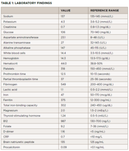
Two weeks later, he presented to the same emergency department with a fever of 103.4º and was admitted for suspected sepsis. He had an extensive evaluation, including blood cultures, a tickborne illness panel, computed tomography (CT) scans of his chest, neck, abdomen and pelvis, and transthoracic cardiac echocardiogram, which showed no acute abnormalities. Laboratory tests and results from that presentation are listed in Table 1.
Additional testing included:
- Serum protein electrophoresis (SPEP): consistent with acute phasereactant, faint restricted-band M spike migrating in the gamma-globulin region;
- Immunofixation: A faint band in IgG kappa could not be ruled out from the serum immunofixation pattern;
- Parvovirus B-19 IgM: negative at 0.1 (cutoff <0.9);
- Parvovirus B-19 IgG: elevated, positive at 4.3; (cutoff <0.9);
- Anti-neutrophilic cytoplasmic antibody (ANCA): negative;
- Hepatitis B and C naive by antibody testing;
- HIV: antibody negative;
- Parvovirus B19 IgM: negative, protein C: normal, protein S: normal;
- Factor 5 Leiden: normal;
- Lupus anticoagulant (dilute Russell viper venom time): 1.19 (cutoff <1.14);
- Anti-centromere antibody: negative;
- Anti-nuclear antibody (ANA): 1:80;
- Double-stranded DNA: negative;
- Rheumatoid factor: negative;
- anti-RNP and anti-Smith antibodies: negative;
- anti-Sjögren’s antibodies: negative;
- Cold agglutinins: negative;
- Urinalysis: normal;
- Legionella antigen: negative;
- Streptococcus pneumonia antigen: negative;
- Epstein Barr virus (EBV) nuclear antibody IgG: positive (cutoff >21.99);
- EBV capsid antibody IgM: negative;
- Cytomegalovirus (CMV) antibody IgM: negative;
- Anaplasma phagocytophilum DNA: not detected;
- Babesi microti DNA: not detected;
- Borrelia miyamotoi DNA: not detected;
- Ehrlichia chaffeensis DNA: negative;
- Lyme disease DNA: not detected.
He was seen by an infectious disease specialist who believed the patient had a viral illness. The patient was discharged.

