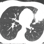
As this is written, it has been more than two years since his presentation to the rheumatologist.
Discussion
The first reported case of PDP was in 1868.1 It most frequently occurs in males, and usually develops in the teenage years, presenting with clubbing, skin thickening and periostosis. The combination of thickened skin and bony enlargement can result in significant thickening of the extremities, which is often a very striking physical finding. The skin thickening can also be very prominent on the forehead and face.2 The digital clubbing, from some reports, occurs in about 89% of cases. There have also been reports of hypertrophy of the eyelids in 30–40% of patients and cutis verticis gyrata in 24% of patients. Cutis verticis gyrata is a thickening of the skin along the scalp that results in an appearance similar to the gyri of the brain.3 Ninety percent of patients tend to have seborrheic dermatitis. Other findings consist of acne, folliculitis, polyarthritis, palpebral ptosis, hyperhidrosis and acro-ostolysis of the long bones.1
Radiographs often reveal diffuse periostosis along the length of bones, including epiphyses in 80–97% of cases, and often with irregular contour.4 The diagnosis of PDP can be based on the presence of at least two of the four criteria set by Borochowitz et al.5:
- History of familial transmission,
- Pachyderma,
- Digital clubbing, and/or
- Skeletal manifestations, such as pain or signs of radiographic periostitis.
PDP has three clinical variants: complete, with pachyderma, clubbing and periostosis; incomplete, with isolated periostosis and limited skin changes; and forme fruste, presenting predominantly with pachyderma, with minimal periostosis.6
Due to the unusual presentation of these patients, many etiologies are often considered at onset, including multiple autoimmune and rheumatic conditions. PDP occurring along with other autoimmune conditions has been reported; one patient had concurrent PDP and limited scleroderma.7 PDP can be distinguished from scleroderma due to periosteal proliferation of the bones and hypertrophic skin changes.
Although the pathogenesis of PDP is still not clearly understood, studies have found genetic abnormalities, with pathogenic theories proposed. A study performed in 2005 reviewed pedigree data published on 68 families with 204 patients.8 The analysis showed evidence of an autosomal dominant mutation in 37 families and suggested the existence of an autosomal recessive form in the remaining families. Despite the incidence of PDP presenting mostly in young men, no X-linked pattern was noted. Theories suggest a potential relation to testosterone effects, such as promoting proliferation. The study did not include genetic testing, only pedigree evaluation.


