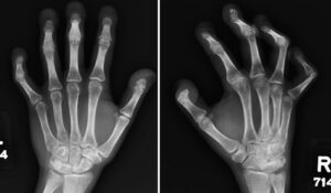
Hajdu-Cheney syndrome (HCS) is a rare connective tissue disease, with fewer than 100 cases reported worldwide.1 Hallmark features include acro-osteolysis (i.e., resorption of the distal phalanges of the hands and feet), osteoporosis, facial dysmorphisms, and craniofacial and dental abnormalities. Patients often have short stature and can have neuroanatomical deformities causing intellectual disabilities. These patients can also have atrioventricular septal defects and polycystic kidneys. HCS is inherited in an autosomal-dominant pattern and is associated with mutations in the NOTCH-2 protein, although the exact mechanism is still under investigation.1 HCS is not only uncommon, but it can present in various ways, making it important to consider in differential diagnoses of several diseases.
We present the case of a young man with upper and lower extremity joint pain and acro-osteolysis presenting for rheumatic evaluation.

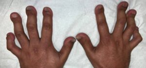
FIGURE 1: Both hands show boutonniere deformities in the third and fourth fingers, with onychorrhexis or longitudinal ridging. (Click to enlarge.)
Case Presentation
Our patient is a 29-year-old man with a past medical history of acro-osteolysis and osteoporosis who presented to our rheumatology clinic with diffuse joint pain. His pain is usually in his right arm in the bicep area and radiates to the forearm. He has associated neuropathy symptoms in both hands. Although his symptoms usually manifest in his right upper extremity, he has had similar symptoms in his left upper and both lower extremities as well. These symptoms occur every few years, persist for a few months and then remit without treatment. He does not have alleviating or aggravating factors and there were no known triggers.
On physical exam, the patient did not have any active synovitis, skin changes or strength deficits. He did have shortened, clubbed digits and flexion deformities of the right fourth and fifth proximal interphalangeal joints. The patient also had a boutonniere deformity in both the third and fourth digits (see Figure 1).
Laboratory studies showed a normal urinalysis and his hemoglobin mildly elevated at 17.7 g/dL (reference range [RR]: 12–15 g/dL). A comprehensive metabolic panel was significant for mildly elevated aspartate transaminase at 43 u/L (RR: 13–39 u/L) and alanine transaminase at 69 u/L (RR: 7/52 u/L). He also had an elevated uric acid at 8.1 mg/dL (RR: 1.5–6.5 mg/dL) and positive anti-mitochondrial M2 antibody at 40.2 u.


