Immune checkpoint inhibitors (ICIs) targeting the cytotoxic T-lymphocyte-associated protein 4 (CTLA-4) or programmed cell death protein 1 (PD-1) axes have revolutionized therapy and improved survival in advanced cancers. However, these immune system modulators also lead to immune-related adverse events (IRAEs).1,2
In clinical trials, IRAEs mainly involved the gastrointestinal tract, skin, endocrine glands, liver and lung, but nearly all organs can be affected. Arthralgias, myalgias and arthritis are the most commonly reported rheumatic IRAEs, but there are case reports of polymyalgia rheumatica (PMR), vasculitis, polymyositis/dermatomyositis, lupus nephritis and scleroderma-like reactions.3,4
In this article, we discuss a case of thyroid myopathy in a patient with diffuse pigmented villonodular synovitis (PVNS) treated with durvalumab (a programmed cell death 1 ligand [PD-1L] inhibitor) and an experimental colony-stimulating factor 1 receptor (CSF-1R) inhibitor, PLX3397. After therapy with the PD-1L inhibitor, the patient developed proximal muscle weakness and elevated muscle enzymes; an elevated thyroid-stimulating hormone (TSH) with undetectable thyroid hormones supported a diagnosis of hypothyroid myopathy. Weakness and muscle enzymes improved after thyroid replacement therapy.
This case highlights the importance of considering endocrinopathies in patients on immunotherapies with myopathy because thyroid dysfunction is one of the most common IRAEs.
Case Presentation
A 54-year-old Hispanic woman with a history of hypothyroidism and recurrent, refractory, diffuse PVNS of her left knee presented to our rheumatology department for progressive proximal muscle pain, weakness and an elevated creatinine kinase (CK) level of 5,635 U/L (reference range [RR]: 40–308 U/L).
She had been diagnosed with diffuse PVNS five years prior to presentation after resection of a left knee mass that demonstrated characteristic oval to epithelioid mononuclear and multinucleated histiocytic-like cells with scattered foamy histiocytes and abundant hemosiderin pigment. She was initially treated with a combination of sirolimus and PLX3397, but the disease recurred after three years, and she had another resection before switching to a second experimental CSF-1R inhibitor, LY3022855, and durvalumab.
Thyroid dysfunction has been reported in up to 10% of patients treated with PD-1/PD-L1 blockade.
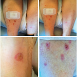
Her second treatment regimen was discontinued after three months due to persistent psoriasiform eruption with inverse features thought to be IRAE secondary to durvalumab. Her rash responded well to one dose of infliximab infusion and prednisone. Her CK was mildly elevated to 631 U/L at that time.
Seven months after discontinuing durvalumab and LY3022855, she was referred to a rheumatologist for worsening muscle pain, proximal weakness and rising muscle enzymes. She had difficulty standing from a seated position and raising her arms above her head, accompanied by shoulder pain. She also complained of decreased grip strength, causing her to frequently drop objects. Her left knee pain got worse when walking upstairs. She had dyspnea on exertion, a brown rash on her extremities and generalized fatigue. She exhibited no dysphagia or dysphonia, had no trouble holding her head up, did not have chest pain, oral or nasal ulcers, Raynaud’s phenomenon or weight loss.


